 Movie
Movie Controller


+ Open data

- Basic information
| Entry | Database: PDB / ID: 10al | |||||||||
|---|---|---|---|---|---|---|---|---|---|---|
| Title | indoleacetate decarboxylase with bound indole-3-acetate | |||||||||
 Components Components | Formate C-acetyltransferase | |||||||||
 Keywords Keywords | LYASE / glycyl radical enzyme / decarboxylase / anaerobic | |||||||||
| Function / homology |  Function and homology information Function and homology informationformate C-acetyltransferase / formate C-acetyltransferase activity / cytosol Similarity search - Function | |||||||||
| Biological species |  Olsenella uli DSM 7084 (bacteria) Olsenella uli DSM 7084 (bacteria) | |||||||||
| Method | ELECTRON MICROSCOPY / single particle reconstruction / cryo EM / Resolution: 2.45 Å | |||||||||
 Authors Authors | Imrich, C.N. / Drennan, C.L. | |||||||||
| Funding support |  United States, 2items United States, 2items
| |||||||||
 Citation Citation | Journal: Proc Natl Acad Sci U S A / Year: 2026 Title: The structural basis of malodorant skatole formation by the glycyl radical enzyme indoleacetate decarboxylase. Authors: Christa N Imrich / Lindsey R F Backman / Abigail P Allworth / Mary C Andorfer / Jared C Paris / Nina M Greeley / Beverly Fu / Emily P Balskus / Catherine L Drennan / Abstract: Glycyl radical enzymes (GREs) catalyze challenging chemical reactions using a posttranslationally installed glycyl radical cofactor. One such enzyme, indoleacetate decarboxylase (IAD), performs the ...Glycyl radical enzymes (GREs) catalyze challenging chemical reactions using a posttranslationally installed glycyl radical cofactor. One such enzyme, indoleacetate decarboxylase (IAD), performs the radical-based decarboxylation of indole-3-acetate (I3A) to form the malodorant molecule skatole. In addition to being an odor nuisance, skatole is a human and livestock lung toxin, a suspected carcinogen, and a mosquito attractant, all of which impact human health, agriculture, food production, and wastewater treatment. Here, we use cryogenic electron microscopy to solve a 2.45-Å resolution structure of IAD from the gut bacterium . We observe IAD in a homotetrameric form with the substrate I3A bound in all four protomers. The positioning of I3A in the active site is unexpected and is more consistent with a Kolbe-type decarboxylation mechanism, i.e., a decarboxylation initiated by a 1-electron oxidation of the carboxylate moiety rather than being initiated by hydrogen atom transfer (HAT). Previously, a high deuterium content in skatole from IAD assays in DO was used to support a HAT mechanism over a Kolbe-type mechanism. However, we show here that deuterium content does not necessarily inform on mechanism as IAD can catalyze the exchange of skatole's 3'-methyl hydrogens postturnover. Structural comparisons show that both IAD and hydroxyphenylacetate decarboxylase display structural features that are not found in other characterized GREs, suggesting that they represent a distinct GRE-subclass. Collectively, these insights will inform IAD inhibitor design aimed at decreasing skatole production. | |||||||||
| History |
|
- Structure visualization
| Structure viewer | Molecule: MolmilJmol/JSmol |
|---|
- Downloads & links
-Download
| PDBx/mmCIF format | 10al.cif.gz | 696.6 KB | Display | PDBx/mmCIF format |
|---|---|---|---|---|
| PDB format | pdb10al.ent.gz | 566.1 KB | Display | PDB format |
| PDBx/mmJSON format | 10al.json.gz | Tree view | PDBx/mmJSON format | |
| Others |  Other downloads Other downloads |
-Validation report
| Arichive directory | https://data.pdbj.org/pub/pdb/validation_reports/0a/10alftp://data.pdbj.org/pub/pdb/validation_reports/0a/10al | HTTPS FTP |
|---|
-Related structure data
| Related structure data |  75028MC M: map data used to model this data C: citing same article ( |
|---|---|
| Similar structure data |
-Links
 PDBj
PDBj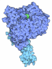
- Assembly
| Deposited unit | 
|
|---|---|
| 1 |
|
-Components
| #1: Protein | Mass: 101384.719 Da / Num. of mol.: 4 Source method: isolated from a genetically manipulated source Details: 6xHis-thrombin cleavage site-IAD / Source: (gene. exp.) Olsenella uli DSM 7084 (bacteria) / Gene: Olsu_0124 / Production host:  Escherichia coli (E. coli) / References: UniProt: E1QXZ2, formate C-acetyltransferase Escherichia coli (E. coli) / References: UniProt: E1QXZ2, formate C-acetyltransferase#2: Chemical | ChemComp-IAC / 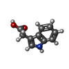 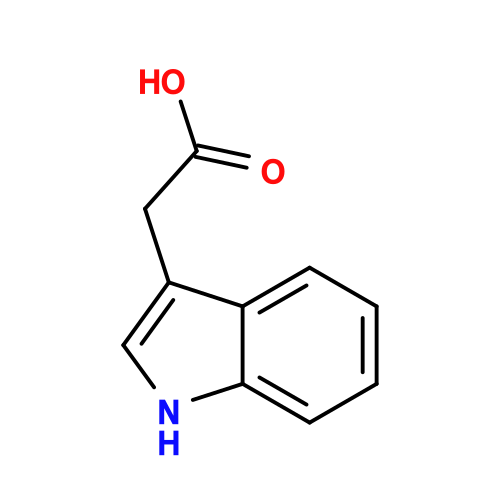 Mass: 175.184 Da / Num. of mol.: 4 / Source method: obtained synthetically / Formula: C10H9NO2 / Feature type: SUBJECT OF INVESTIGATION / Comment: hormone*YM Mass: 175.184 Da / Num. of mol.: 4 / Source method: obtained synthetically / Formula: C10H9NO2 / Feature type: SUBJECT OF INVESTIGATION / Comment: hormone*YM#3: Water | ChemComp-HOH / |  Mass: 18.015 Da / Num. of mol.: 685 / Source method: isolated from a natural source / Formula: H2O Mass: 18.015 Da / Num. of mol.: 685 / Source method: isolated from a natural source / Formula: H2OHas ligand of interest | Y | Has protein modification | N | |
|---|
-Experimental details
-Experiment
| Experiment | Method: ELECTRON MICROSCOPY |
|---|---|
| EM experiment | Aggregation state: PARTICLE / 3D reconstruction method: single particle reconstruction |
- Sample preparation
| Component | Name: homotetramer of indoleacetate decarboxylase with bound indole-3-acetate Type: COMPLEX / Entity ID: #1 / Source: RECOMBINANT | |||||||||||||||||||||||||
|---|---|---|---|---|---|---|---|---|---|---|---|---|---|---|---|---|---|---|---|---|---|---|---|---|---|---|
| Molecular weight | Value: 0.101 MDa / Experimental value: NO | |||||||||||||||||||||||||
| Source (natural) | Organism: Olsenella uli DSM 7084 (bacteria) | |||||||||||||||||||||||||
| Source (recombinant) | Organism: Escherichia coli (E. coli) / Strain: T7 Express / Plasmid: pET28a | |||||||||||||||||||||||||
| Buffer solution | pH: 7.5 | |||||||||||||||||||||||||
| Buffer component |
| |||||||||||||||||||||||||
| Specimen | Conc.: 10 mg/ml / Embedding applied: NO / Shadowing applied: NO / Staining applied: NO / Vitrification applied: YES / Details: isolated dimer by gel filtration chromatography | |||||||||||||||||||||||||
| Specimen support | Grid material: COPPER / Grid mesh size: 300 divisions/in. / Grid type: Quantifoil Active R1.2/0.8 | |||||||||||||||||||||||||
| Vitrification | Instrument: SPT LABTECH CHAMELEON / Cryogen name: ETHANE / Humidity: 81 % / Chamber temperature: 297.35 K / Details: plunge time 155 ms |
- Electron microscopy imaging
| Experimental equipment |  Model: Titan Krios / Image courtesy: FEI Company |
|---|---|
| Microscopy | Model: TFS KRIOS |
| Electron gun | Electron source:  FIELD EMISSION GUN / Accelerating voltage: 300 kV / Illumination mode: FLOOD BEAM FIELD EMISSION GUN / Accelerating voltage: 300 kV / Illumination mode: FLOOD BEAM |
| Electron lens | Mode: BRIGHT FIELD / Nominal magnification: 105000 X / Nominal defocus max: 2000 nm / Nominal defocus min: 200 nm / Cs: 2.7 mm |
| Specimen holder | Cryogen: NITROGEN / Specimen holder model: FEI TITAN KRIOS AUTOGRID HOLDER |
| Image recording | Average exposure time: 1.9 sec. / Electron dose: 52.07 e/Å2 / Film or detector model: GATAN K3 BIOQUANTUM (6k x 4k) / Num. of grids imaged: 1 / Num. of real images: 8927 |
- Processing
| EM software |
| ||||||||||||||||||||||||||||||||
|---|---|---|---|---|---|---|---|---|---|---|---|---|---|---|---|---|---|---|---|---|---|---|---|---|---|---|---|---|---|---|---|---|---|
| CTF correction | Type: PHASE FLIPPING AND AMPLITUDE CORRECTION | ||||||||||||||||||||||||||||||||
| Particle selection | Num. of particles selected: 2743992 | ||||||||||||||||||||||||||||||||
| 3D reconstruction | Resolution: 2.45 Å / Resolution method: FSC 0.143 CUT-OFF / Num. of particles: 330270 / Symmetry type: POINT | ||||||||||||||||||||||||||||||||
| Atomic model building | Protocol: RIGID BODY FIT / Space: REAL | ||||||||||||||||||||||||||||||||
| Atomic model building | Chain residue range: 1-878 / Source name: AlphaFold / Type: in silico model |
