 Movie
Movie Controller


+ Open data

- Basic information
| Entry |  | |||||||||
|---|---|---|---|---|---|---|---|---|---|---|
| Title | indoleacetate decarboxylase with bound indole-3-acetate | |||||||||
 Map data Map data | OuIAD tetramer map | |||||||||
 Sample Sample |
| |||||||||
 Keywords Keywords | glycyl radical enzyme / decarboxylase / anaerobic / LYASE | |||||||||
| Function / homology |  Function and homology information Function and homology informationformate C-acetyltransferase / formate C-acetyltransferase activity / cytosol Similarity search - Function | |||||||||
| Biological species |  Olsenella uli DSM 7084 (bacteria) Olsenella uli DSM 7084 (bacteria) | |||||||||
| Method | single particle reconstruction / cryo EM / Resolution: 2.45 Å | |||||||||
 Authors Authors | Imrich CN / Drennan CL | |||||||||
| Funding support |  United States, 2 items United States, 2 items
| |||||||||
 Citation Citation | Journal: Proc Natl Acad Sci U S A / Year: 2026 Title: The structural basis of malodorant skatole formation by the glycyl radical enzyme indoleacetate decarboxylase. Authors: Christa N Imrich / Lindsey R F Backman / Abigail P Allworth / Mary C Andorfer / Jared C Paris / Nina M Greeley / Beverly Fu / Emily P Balskus / Catherine L Drennan / Abstract: Glycyl radical enzymes (GREs) catalyze challenging chemical reactions using a posttranslationally installed glycyl radical cofactor. One such enzyme, indoleacetate decarboxylase (IAD), performs the ...Glycyl radical enzymes (GREs) catalyze challenging chemical reactions using a posttranslationally installed glycyl radical cofactor. One such enzyme, indoleacetate decarboxylase (IAD), performs the radical-based decarboxylation of indole-3-acetate (I3A) to form the malodorant molecule skatole. In addition to being an odor nuisance, skatole is a human and livestock lung toxin, a suspected carcinogen, and a mosquito attractant, all of which impact human health, agriculture, food production, and wastewater treatment. Here, we use cryogenic electron microscopy to solve a 2.45-Å resolution structure of IAD from the gut bacterium . We observe IAD in a homotetrameric form with the substrate I3A bound in all four protomers. The positioning of I3A in the active site is unexpected and is more consistent with a Kolbe-type decarboxylation mechanism, i.e., a decarboxylation initiated by a 1-electron oxidation of the carboxylate moiety rather than being initiated by hydrogen atom transfer (HAT). Previously, a high deuterium content in skatole from IAD assays in DO was used to support a HAT mechanism over a Kolbe-type mechanism. However, we show here that deuterium content does not necessarily inform on mechanism as IAD can catalyze the exchange of skatole's 3'-methyl hydrogens postturnover. Structural comparisons show that both IAD and hydroxyphenylacetate decarboxylase display structural features that are not found in other characterized GREs, suggesting that they represent a distinct GRE-subclass. Collectively, these insights will inform IAD inhibitor design aimed at decreasing skatole production. | |||||||||
| History |
|
- Structure visualization
| Supplemental images |
|---|

- Downloads & links
-EMDB archive
| Map data | emd_75028.map.gz | 17.3 MB | EMDB map data format | |
|---|---|---|---|---|
| Header (meta data) | emd-75028-v30.xmlemd-75028.xml | 23.5 KB 23.5 KB | Display Display | EMDB header |
| FSC (resolution estimation) | emd_75028_fsc.xml | 12.8 KB | Display | FSC data file |
| Images |  emd_75028.png emd_75028.png | 117.5 KB | ||
| Filedesc metadata | emd-75028.cif.gz | 7.3 KB | ||
| Others | emd_75028_half_map_1.map.gzemd_75028_half_map_2.map.gz | 141.3 MB 141.3 MB | ||
| Archive directory |  https://data.pdbj.org/pub/emdb/structures/EMD-75028ftp://data.pdbj.org/pub/emdb/structures/EMD-75028 https://data.pdbj.org/pub/emdb/structures/EMD-75028ftp://data.pdbj.org/pub/emdb/structures/EMD-75028 | HTTPS FTP |
-Related structure data
| Related structure data |  10alMC M: atomic model generated by this map C: citing same article ( |
|---|---|
| Similar structure data |
-Links
| EMDB pages | EMDB (EBI/PDBe) / EMDataResource |
|---|
-Map
| File | Download / File: emd_75028.map.gz / Format: CCP4 / Size: 178 MB / Type: IMAGE STORED AS FLOATING POINT NUMBER (4 BYTES) | ||||||||||||||||||||||||||||||||||||
|---|---|---|---|---|---|---|---|---|---|---|---|---|---|---|---|---|---|---|---|---|---|---|---|---|---|---|---|---|---|---|---|---|---|---|---|---|---|
| Annotation | OuIAD tetramer map | ||||||||||||||||||||||||||||||||||||
| Projections & slices | Image control
Images are generated by Spider. | ||||||||||||||||||||||||||||||||||||
| Voxel size | X=Y=Z: 0.822 Å | ||||||||||||||||||||||||||||||||||||
| Density |
| ||||||||||||||||||||||||||||||||||||
| Symmetry | Space group: 1 | ||||||||||||||||||||||||||||||||||||
| Details | EMDB XML:
|
 Z (Sec.)
 Y (Row.)
 X (Col.)


-Supplemental data
-Half map: Half map 2
| File | emd_75028_half_map_1.map | ||||||||||||
|---|---|---|---|---|---|---|---|---|---|---|---|---|---|
| Annotation | Half map 2 | ||||||||||||
| Projections & Slices |
| ||||||||||||
| Density Histograms |


-Half map: Half map 1
| File | emd_75028_half_map_2.map | ||||||||||||
|---|---|---|---|---|---|---|---|---|---|---|---|---|---|
| Annotation | Half map 1 | ||||||||||||
| Projections & Slices |
| ||||||||||||
| Density Histograms |


- Sample components
-Entire : homotetramer of indoleacetate decarboxylase with bound indole-3-a...
| Entire | Name: homotetramer of indoleacetate decarboxylase with bound indole-3-acetate |
|---|---|
| Components |
|
-Supramolecule #1: homotetramer of indoleacetate decarboxylase with bound indole-3-a...
| Supramolecule | Name: homotetramer of indoleacetate decarboxylase with bound indole-3-acetate type: complex / ID: 1 / Parent: 0 / Macromolecule list: #1 |
|---|---|
| Source (natural) | Organism: Olsenella uli DSM 7084 (bacteria) |
| Molecular weight | Theoretical: 101 KDa |
-Macromolecule #1: Formate C-acetyltransferase
| Macromolecule | Name: Formate C-acetyltransferase / type: protein_or_peptide / ID: 1 / Details: 6xHis-thrombin cleavage site-IAD / Number of copies: 4 / Enantiomer: LEVO / EC number: formate C-acetyltransferase |
|---|---|
| Source (natural) | Organism: Olsenella uli DSM 7084 (bacteria) |
| Molecular weight | Theoretical: 101.384719 KDa |
| Recombinant expression | Organism:  Escherichia coli (E. coli) Escherichia coli (E. coli) |
| Sequence | String: MGSSHHHHHH SSGLVPRGSH MEESLVLEML QTGKTATWPA KNQLQESGEV VDKEVKGTPS TDRTRRMKER FMNAKCKMDM EAPIAYTKA WREHEGKPLY VRRGLAYKYM LEHLTPAIRE DELITMSKTR YDRGATQVPQ FATDFMISFL TQAEDQKEEA K LYSVEGKD ...String: MGSSHHHHHH SSGLVPRGSH MEESLVLEML QTGKTATWPA KNQLQESGEV VDKEVKGTPS TDRTRRMKER FMNAKCKMDM EAPIAYTKA WREHEGKPLY VRRGLAYKYM LEHLTPAIRE DELITMSKTR YDRGATQVPQ FATDFMISFL TQAEDQKEEA K LYSVEGKD EAHTVEEEGW TKVGQLFSIR EEEVKPMLEV LEYWKTRCVE NVSDDWMKTS FPYYQDYVNA KKVGLFPGSG LH AGCDGRW IPAYDVALGG LNRVIEECRE KIEKTVVTTK EVADKVFFWQ GCIYACEGAI AWAHNYAVEA RRLAETAVEP RKT ELLEMA ERLDRVPAEA PRNFMEAVQA LWTTQILVIS DSLALGVSPG RWGKFLEPYY EKDLAEGRIT KGQALEVMEL LRIK FSTEE YITPSLWAAM ASSNSFMNLA VGGLDPKTGK CTDNEIEDLI LEAGINMPTP QPTLSILLSD KTTDHLAIKA AECTK AGNG YPAWFNYDMM VQHNLAMYRD EGITLEDARN CALSGCVENG LAGTGHPIAH PAFYNEGKTI ELACNEGVDP RTGIKV MDG IVPIKTYEDL WDNFIKIREH FMHVYMQYWN EVVACQRDIH PKIMGSVLMH DCIESGRPVD NLGCRYNGSV TLLDSGT VN VVNGLAAIKK LVFEDHKYTW DEFKEAMDNN FGFVLGAEKG NFSMLNQEID PEKHMKYAHI HRDVLNAPKF GNDDDFVD D IFVDLWHDYD RVTASETTYN GYRWITAALS ISAHGPHGRV TGATPDGRLS GVTLCDGILS ASPGTDVNGP IALIRSGVK LDPTEFASVQ LNMKFHPTAI RGDEGSRNFV DFIHSYFQMG GYHVQFNIVD SKMLRDAQDH PQNYRDLMVR VAGFSAYWNE LGKPIQDEV IARTEYDAL UniProtKB: Formate C-acetyltransferase |
-Macromolecule #2: 1H-INDOL-3-YLACETIC ACID
| Macromolecule | Name: 1H-INDOL-3-YLACETIC ACID / type: ligand / ID: 2 / Number of copies: 4 / Formula: IAC |
|---|---|
| Molecular weight | Theoretical: 175.184 Da |
| Chemical component information | 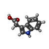 ChemComp-IAC: |
-Macromolecule #3: water
| Macromolecule | Name: water / type: ligand / ID: 3 / Number of copies: 685 / Formula: HOH |
|---|---|
| Molecular weight | Theoretical: 18.015 Da |
| Chemical component information |  ChemComp-HOH: |
-Experimental details
-Structure determination
| Method | cryo EM |
|---|---|
 Processing Processing | single particle reconstruction |
| Aggregation state | particle |
-Sample preparation
| Concentration | 10 mg/mL | |||||||||||||||
|---|---|---|---|---|---|---|---|---|---|---|---|---|---|---|---|---|
| Buffer | pH: 7.5 Component:
| |||||||||||||||
| Grid | Model: Quantifoil Active R1.2/0.8 / Material: COPPER / Mesh: 300 / Support film - Material: CARBON / Support film - topology: HOLEY / Pretreatment - Type: GLOW DISCHARGE / Pretreatment - Time: 240 sec. | |||||||||||||||
| Vitrification | Cryogen name: ETHANE / Chamber humidity: 81 % / Chamber temperature: 297.35 K / Instrument: SPT LABTECH CHAMELEON / Details: plunge time 155 ms. | |||||||||||||||
| Details | isolated dimer by gel filtration chromatography |
- Electron microscopy
| Microscope | TFS KRIOS |
|---|---|
| Image recording | Film or detector model: GATAN K3 BIOQUANTUM (6k x 4k) / Number grids imaged: 1 / Number real images: 8927 / Average exposure time: 1.9 sec. / Average electron dose: 52.07 e/Å2 |
| Electron beam | Acceleration voltage: 300 kV / Electron source:  FIELD EMISSION GUN FIELD EMISSION GUN |
| Electron optics | Illumination mode: FLOOD BEAM / Imaging mode: BRIGHT FIELD / Cs: 2.7 mm / Nominal defocus max: 2.0 µm / Nominal defocus min: 0.2 µm / Nominal magnification: 105000 |
| Sample stage | Specimen holder model: FEI TITAN KRIOS AUTOGRID HOLDER / Cooling holder cryogen: NITROGEN |
| Experimental equipment |  Model: Titan Krios / Image courtesy: FEI Company |
+Image processing

-Atomic model buiding 1
| Initial model | Chain - Residue range: 1-878 / Chain - Source name: AlphaFold / Chain - Initial model type: in silico model |
|---|---|
| Refinement | Space: REAL / Protocol: RIGID BODY FIT |
| Output model | PDB-10al: |
