[English] 日本語
 Yorodumi
- EMDB-9854: Structure of the green algal photosystem I supercomplex with ligh... -
+ Open data
- Basic information
| Entry | Database: EMDB / ID: EMD-9854 | ||||||||||||
|---|---|---|---|---|---|---|---|---|---|---|---|---|---|
| Title | Structure of the green algal photosystem I supercomplex with light-harvesting complex I | ||||||||||||
 Map data Map data | |||||||||||||
 Sample Sample |
| ||||||||||||
 Keywords Keywords | photosystem membrane protein / PHOTOSYNTHESIS | ||||||||||||
| Function / homology |  Function and homology information Function and homology informationphotosynthesis, light harvesting in photosystem I / photosynthesis, light harvesting / chloroplast thylakoid lumen / photosystem I reaction center / photosystem I / photosynthetic electron transport in photosystem I / photosystem I / photosystem II / chlorophyll binding / chloroplast thylakoid membrane ...photosynthesis, light harvesting in photosystem I / photosynthesis, light harvesting / chloroplast thylakoid lumen / photosystem I reaction center / photosystem I / photosynthetic electron transport in photosystem I / photosystem I / photosystem II / chlorophyll binding / chloroplast thylakoid membrane / response to light stimulus / photosynthesis / 4 iron, 4 sulfur cluster binding / electron transfer activity / oxidoreductase activity / magnesium ion binding / metal ion binding Similarity search - Function | ||||||||||||
| Biological species |  | ||||||||||||
| Method | single particle reconstruction / cryo EM / Resolution: 2.9 Å | ||||||||||||
 Authors Authors | Suga M / Miyazaki N | ||||||||||||
| Funding support |  Japan, 3 items Japan, 3 items
| ||||||||||||
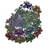
 Citation Citation |  Journal: Nat Plants / Year: 2019 Journal: Nat Plants / Year: 2019Title: Structure of the green algal photosystem I supercomplex with a decameric light-harvesting complex I. Authors: Michihiro Suga / Shin-Ichiro Ozawa / Kaori Yoshida-Motomura / Fusamichi Akita / Naoyuki Miyazaki / Yuichiro Takahashi /  Abstract: In plants and green algae, the core of photosystem I (PSI) is surrounded by a peripheral antenna system consisting of light-harvesting complex I (LHCI). Here we report the cryo-electron ...In plants and green algae, the core of photosystem I (PSI) is surrounded by a peripheral antenna system consisting of light-harvesting complex I (LHCI). Here we report the cryo-electron microscopic structure of the PSI-LHCI supercomplex from the green alga Chlamydomonas reinhardtii. The structure reveals that eight Lhca proteins form two tetrameric LHCI belts attached to the PsaF side while the other two Lhca proteins form an additional Lhca2/Lhca9 heterodimer attached to the opposite side. The spatial arrangement of light-harvesting pigments reveals that Chlorophylls b are more abundant in the outer LHCI belt than in the inner LHCI belt and are absent from the core, thereby providing the downhill energy transfer pathways to the PSI core. PSI-LHCI is complexed with a plastocyanin on the patch of lysine residues of PsaF at the luminal side. The assembly provides a structural basis for understanding the mechanism of light-harvesting, excitation energy transfer of the PSI-LHCI supercomplex and electron transfer with plastocyanin. | ||||||||||||
| History |
|
- Structure visualization
| Movie |
 Movie viewer Movie viewer |
|---|---|
| Structure viewer | EM map:  SurfView SurfView Molmil Molmil Jmol/JSmol Jmol/JSmol |
| Supplemental images |
- Downloads & links
-EMDB archive
| Map data |  emd_9854.map.gz emd_9854.map.gz | 13.3 MB |  EMDB map data format EMDB map data format | |
|---|---|---|---|---|
| Header (meta data) |  emd-9854-v30.xml emd-9854-v30.xml emd-9854.xml emd-9854.xml | 43.8 KB 43.8 KB | Display Display |  EMDB header EMDB header |
| Images |  emd_9854.png emd_9854.png | 64.4 KB | ||
| Filedesc metadata |  emd-9854.cif.gz emd-9854.cif.gz | 10.6 KB | ||
| Archive directory |  http://ftp.pdbj.org/pub/emdb/structures/EMD-9854 http://ftp.pdbj.org/pub/emdb/structures/EMD-9854 ftp://ftp.pdbj.org/pub/emdb/structures/EMD-9854 ftp://ftp.pdbj.org/pub/emdb/structures/EMD-9854 | HTTPS FTP |
-Related structure data
| Related structure data |  6jo6MC  9853C  9855C  9856C  6jo5C M: atomic model generated by this map C: citing same article ( |
|---|---|
| Similar structure data | |
| EM raw data |  EMPIAR-10314 (Title: Structure of the green algal photosystem I supercomplex with a decameric light-harvesting complex I. EMPIAR-10314 (Title: Structure of the green algal photosystem I supercomplex with a decameric light-harvesting complex I.Data size: 8.0 TB Data #1: Raw multiframe micrographs [micrographs - multiframe]) |
- Links
Links
| EMDB pages |  EMDB (EBI/PDBe) / EMDB (EBI/PDBe) /  EMDataResource EMDataResource |
|---|---|
| Related items in Molecule of the Month |
- Map
Map
| File |  Download / File: emd_9854.map.gz / Format: CCP4 / Size: 125 MB / Type: IMAGE STORED AS FLOATING POINT NUMBER (4 BYTES) Download / File: emd_9854.map.gz / Format: CCP4 / Size: 125 MB / Type: IMAGE STORED AS FLOATING POINT NUMBER (4 BYTES) | ||||||||||||||||||||||||||||||||||||||||||||||||||||||||||||||||||||
|---|---|---|---|---|---|---|---|---|---|---|---|---|---|---|---|---|---|---|---|---|---|---|---|---|---|---|---|---|---|---|---|---|---|---|---|---|---|---|---|---|---|---|---|---|---|---|---|---|---|---|---|---|---|---|---|---|---|---|---|---|---|---|---|---|---|---|---|---|---|
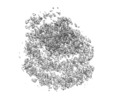
| Projections & slices | Image control
Images are generated by Spider. | ||||||||||||||||||||||||||||||||||||||||||||||||||||||||||||||||||||
| Voxel size | X=Y=Z: 1.12 Å | ||||||||||||||||||||||||||||||||||||||||||||||||||||||||||||||||||||
| Density |
| ||||||||||||||||||||||||||||||||||||||||||||||||||||||||||||||||||||
| Symmetry | Space group: 1 | ||||||||||||||||||||||||||||||||||||||||||||||||||||||||||||||||||||
| Details | EMDB XML:
CCP4 map header:
| ||||||||||||||||||||||||||||||||||||||||||||||||||||||||||||||||||||
-Supplemental data
- Sample components
+Entire : Photosystem I - Light Harvesting Complex I supercomplex
+Supramolecule #1: Photosystem I - Light Harvesting Complex I supercomplex
+Macromolecule #1: Photosystem I P700 chlorophyll a apoprotein A1
+Macromolecule #2: Photosystem I P700 chlorophyll a apoprotein A2
+Macromolecule #3: Photosystem I iron-sulfur center
+Macromolecule #4: Photosystem I reaction center subunit II, chloroplastic
+Macromolecule #5: Photosystem I reaction center subunit IV, chloroplastic
+Macromolecule #6: Photosystem I reaction center subunit F, Photosystem I reaction c...
+Macromolecule #7: Photosystem I reaction center subunit V, chloroplastic
+Macromolecule #8: Photosystem I reaction center subunit VIII
+Macromolecule #9: Photosystem I reaction center subunit IX
+Macromolecule #10: Photosystem I reaction center subunit psaK, chloroplastic
+Macromolecule #11: Photosystem I reaction center subunit XI
+Macromolecule #12: Chlorophyll a-b binding protein, chloroplastic
+Macromolecule #13: Chlorophyll a-b binding protein, chloroplastic
+Macromolecule #14: Chlorophyll a-b binding protein, chloroplastic
+Macromolecule #15: Chlorophyll a-b binding protein, chloroplastic
+Macromolecule #16: Chlorophyll a-b binding protein, chloroplastic
+Macromolecule #17: Chlorophyll a-b binding protein, chloroplastic
+Macromolecule #18: Chlorophyll a-b binding protein, chloroplastic
+Macromolecule #19: CHLOROPHYLL A ISOMER
+Macromolecule #20: CHLOROPHYLL A
+Macromolecule #21: PHYLLOQUINONE
+Macromolecule #22: 1,2-DIPALMITOYL-PHOSPHATIDYL-GLYCEROLE
+Macromolecule #23: BETA-CAROTENE
+Macromolecule #24: IRON/SULFUR CLUSTER
+Macromolecule #25: DIGALACTOSYL DIACYL GLYCEROL (DGDG)
+Macromolecule #26: 1,2-DISTEAROYL-MONOGALACTOSYL-DIGLYCERIDE
+Macromolecule #27: CHLOROPHYLL B
+Macromolecule #28: (3R,3'R,6S)-4,5-DIDEHYDRO-5,6-DIHYDRO-BETA,BETA-CAROTENE-3,3'-DIOL
-Experimental details
-Structure determination
| Method | cryo EM |
|---|---|
 Processing Processing | single particle reconstruction |
| Aggregation state | particle |
- Sample preparation
| Buffer | pH: 7.5 |
|---|---|
| Grid | Model: Quantifoil R1.2/1.3 / Material: MOLYBDENUM / Mesh: 300 / Support film - Material: CARBON / Support film - topology: CONTINUOUS / Pretreatment - Type: GLOW DISCHARGE / Pretreatment - Time: 30 sec. |
| Vitrification | Cryogen name: ETHANE |
- Electron microscopy
| Microscope | FEI TITAN KRIOS |
|---|---|
| Image recording | Film or detector model: FEI FALCON III (4k x 4k) / Number grids imaged: 2 / Number real images: 9801 / Average exposure time: 2.5 sec. / Average electron dose: 50.0 e/Å2 |
| Electron beam | Acceleration voltage: 300 kV / Electron source:  FIELD EMISSION GUN FIELD EMISSION GUN |
| Electron optics | C2 aperture diameter: 100.0 µm / Illumination mode: FLOOD BEAM / Imaging mode: BRIGHT FIELD / Nominal defocus max: 3.75 µm / Nominal defocus min: 1.6 µm |
| Sample stage | Specimen holder model: FEI TITAN KRIOS AUTOGRID HOLDER / Cooling holder cryogen: NITROGEN |
| Experimental equipment |  Model: Titan Krios / Image courtesy: FEI Company |
 Movie
Movie Controller






 Z (Sec.)
 Y (Row.)
 X (Col.)


