National Institutes of Health/National Institute of General Medical Sciences (NIH/NIGMS)
R01 GM110185
United States
National Science Foundation (NSF, United States)
DBI-0939454
United States
Citation
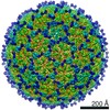
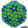
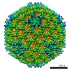
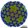
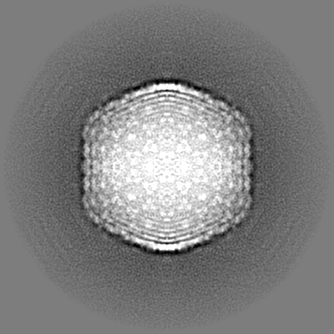
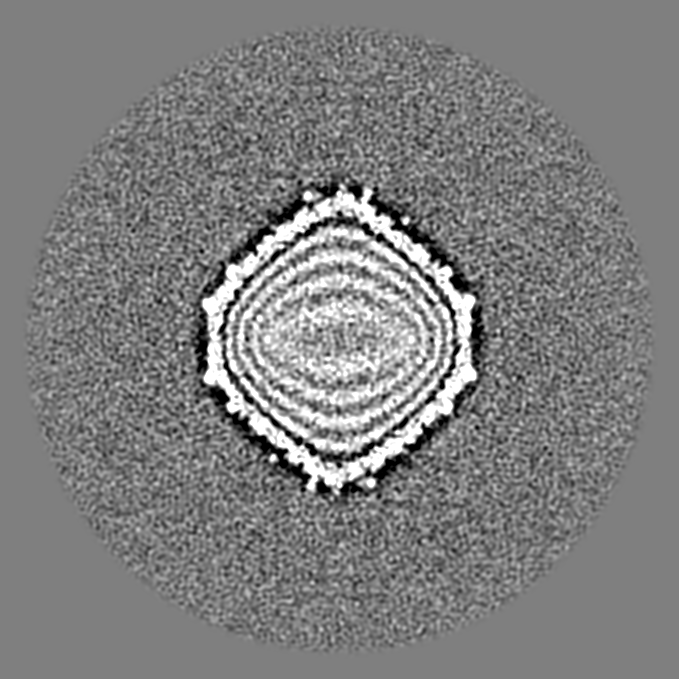
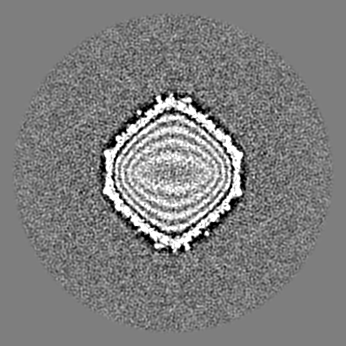
Journal: J Virol / Year: 2018 Title: Shigella Phages Isolated during a Dysentery Outbreak Reveal Uncommon Structures and Broad Species Diversity. Authors: Sarah M Doore / Jason R Schrad / William F Dean / John A Dover / Kristin N Parent / Abstract: In 2016, Michigan experienced the largest outbreak of shigellosis, a type of bacillary dysentery caused by spp., since 1988. Following this outbreak, we isolated 16 novel -infecting bacteriophages ...In 2016, Michigan experienced the largest outbreak of shigellosis, a type of bacillary dysentery caused by spp., since 1988. Following this outbreak, we isolated 16 novel -infecting bacteriophages (viruses that infect bacteria) from environmental water sources. Most well-known bacteriophages infect the common laboratory species and , and these phages have built the foundation of molecular and bacteriophage biology. Until now, comparatively few bacteriophages were known to infect spp., which are close relatives of We present a comprehensive analysis of these phages' host ranges, genomes, and structures, revealing genome sizes and capsid properties that are shared by very few previously described phages. After sequencing, a majority of the phages were found to have genomes of an uncommon size, shared by only 2% of all reported phage genomes. To investigate the structural implications of this unusual genome size, we used cryo-electron microscopy to resolve their capsid structures. We determined that these bacteriophage capsids have similarly uncommon geometry. Only two other viruses with this capsid structure have been described. Since most well-known bacteriophages infect or , our understanding of bacteriophages has been limited to a subset of well-described systems. Continuing to isolate phages using nontraditional strains of bacteria can fill gaps that currently exist in bacteriophage biology. In addition, the prevalence of phages during a shigellosis outbreak may suggest a potential impact of human health epidemics on local microbial communities. spp. bacteria are causative agents of dysentery and affect more than 164 million people worldwide every year. Despite the need to combat antibiotic-resistant strains, relatively few -infecting bacteriophages have been described. By specifically looking for -infecting phages, this work has identified new isolates that (i) may be useful to combat infections and (ii) fill gaps in our knowledge of bacteriophage biology. The rare qualities of these new isolates emphasize the importance of isolating phages on "nontraditional" laboratory strains of bacteria to more fully understand both the basic biology and diversity of bacteriophages.
History
Deposition
Aug 1, 2017
-
Header (metadata) release
Jan 17, 2018
-
Map release
Jan 17, 2018
-
Update
Dec 25, 2019
-
Current status
Dec 25, 2019
Processing site: RCSB / Status: Released
-
Structure visualization
Movie
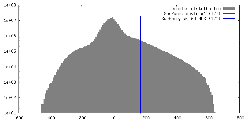
Surface view with section colored by density value
In the structure databanks used in Yorodumi, some data are registered as the other names, "COVID-19 virus" and "2019-nCoV". Here are the details of the virus and the list of structure data.
Jan 31, 2019. EMDB accession codes are about to change! (news from PDBe EMDB page)
EMDB accession codes are about to change! (news from PDBe EMDB page)
The allocation of 4 digits for EMDB accession codes will soon come to an end. Whilst these codes will remain in use, new EMDB accession codes will include an additional digit and will expand incrementally as the available range of codes is exhausted. The current 4-digit format prefixed with “EMD-” (i.e. EMD-XXXX) will advance to a 5-digit format (i.e. EMD-XXXXX), and so on. It is currently estimated that the 4-digit codes will be depleted around Spring 2019, at which point the 5-digit format will come into force.
The EM Navigator/Yorodumi systems omit the EMD- prefix.
Related info.:Q: What is EMD? / ID/Accession-code notation in Yorodumi/EM Navigator
Yorodumi is a browser for structure data from EMDB, PDB, SASBDB, etc.
This page is also the successor to EM Navigator detail page, and also detail information page/front-end page for Omokage search.
The word "yorodu" (or yorozu) is an old Japanese word meaning "ten thousand". "mi" (miru) is to see.
Related info.:EMDB / PDB / SASBDB / Comparison of 3 databanks / Yorodumi Search / Aug 31, 2016. New EM Navigator & Yorodumi / Yorodumi Papers / Jmol/JSmol / Function and homology information / Changes in new EM Navigator and Yorodumi
 Movie
Movie Controller


 Open data
 Basic information
 Map data
 Sample
 Bacteriophage Sf14 (virus)
 Authors
 United States, 2 items
  Citation
 Structure visualization
 Movie viewer
 Downloads & links
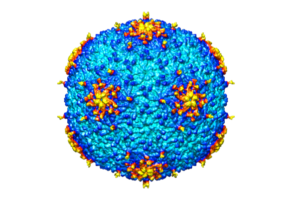 emd_8869.png
 http://ftp.pdbj.org/pub/emdb/structures/EMD-8869

 Z (Sec.)
 Y (Row.)
 X (Col.)


 Sample components
 Shigella flexneri (bacteria) / Strain: PE577
 Processing
 Electron microscopy
 FIELD EMISSION GUN