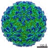
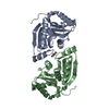
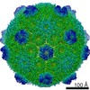
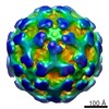
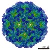
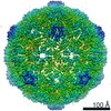
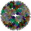
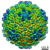
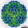
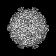
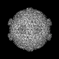
Journal: Science / Year: 2017 Title: Assembly principles and structure of a 6.5-MDa bacterial microcompartment shell. Authors: Markus Sutter / Basil Greber / Clement Aussignargues / Cheryl A Kerfeld / Abstract: Many bacteria contain primitive organelles composed entirely of protein. These bacterial microcompartments share a common architecture of an enzymatic core encapsulated in a selectively permeable ...Many bacteria contain primitive organelles composed entirely of protein. These bacterial microcompartments share a common architecture of an enzymatic core encapsulated in a selectively permeable protein shell; prominent examples include the carboxysome for CO fixation and catabolic microcompartments found in many pathogenic microbes. The shell sequesters enzymatic reactions from the cytosol, analogous to the lipid-based membrane of eukaryotic organelles. Despite available structural information for single building blocks, the principles of shell assembly have remained elusive. We present the crystal structure of an intact shell from , revealing the basic principles of bacterial microcompartment shell construction. Given the conservation among shell proteins of all bacterial microcompartments, these principles apply to functionally diverse organelles and can inform the design and engineering of shells with new functionalities.
History
Deposition
May 26, 2017
-
Header (metadata) release
Jun 14, 2017
-
Map release
Jul 5, 2017
-
Update
Jun 13, 2018
-
Current status
Jun 13, 2018
Processing site: RCSB / Status: Released
-
Structure visualization
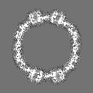
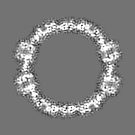
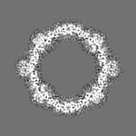
Movie
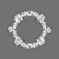
Surface view with section colored by density value
Organism: Escherichia coli (E. coli) / Recombinant strain: BL21 (DE3)
Molecular weight
Theoretical: 6.5 MDa
-
Experimental details
-
Structure determination
Method
cryo EM
Processing
single particle reconstruction
Aggregation state
particle
-
Sample preparation
Concentration
3 mg/mL
Buffer
pH: 7.4 Component:
Concentration
Name
Formula
20.0 mM
Tris-HCl
50.0 mM
Sodium chloride
NaCl
0.01 %
NP-40 substitute
Grid
Model: Protochips CF-1.2/1.3 / Material: COPPER / Mesh: 400 / Support film - #0 - Film type ID: 1 / Support film - #0 - Material: CARBON / Support film - #0 - topology: HOLEY / Support film - #1 - Film type ID: 2 / Support film - #1 - Material: CARBON / Support film - #1 - topology: CONTINUOUS / Pretreatment - Type: PLASMA CLEANING
Vitrification
Cryogen name: ETHANE / Chamber humidity: 100 % / Chamber temperature: 277 K / Instrument: FEI VITROBOT MARK IV / Details: Incubation on grid for 5-7 sec before plunging.
-
Electron microscopy
Microscope
FEI TECNAI F20
Image recording
Film or detector model: GATAN ULTRASCAN 4000 (4k x 4k) / Digitization - Dimensions - Width: 4096 pixel / Digitization - Dimensions - Height: 4096 pixel / Digitization - Sampling interval: 15.0 µm / Number grids imaged: 1 / Number real images: 90 / Average exposure time: 0.4 sec. / Average electron dose: 25.0 e/Å2 / Details: Semi-automated collection using LEGINON
Electron beam
Acceleration voltage: 120 kV / Electron source: FIELD EMISSION GUN
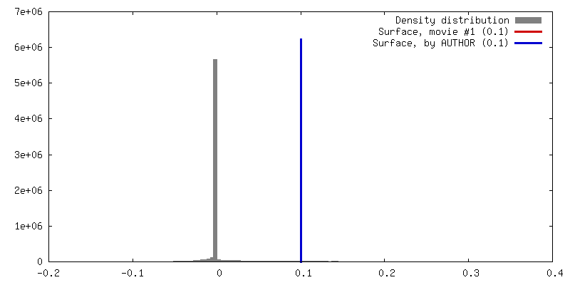
Details: EMD-3351 was scaled to match the size of the BMC shell and low-pass filtered to avoid model bias.
Final reconstruction
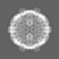
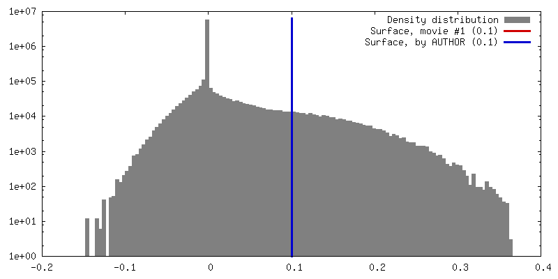
Number classes used: 2 / Applied symmetry - Point group: I (icosahedral) / Resolution.type: BY AUTHOR / Resolution: 8.7 Å / Resolution method: FSC 0.143 CUT-OFF / Software - Name: RELION (ver. 1.4) / Details: Icosahedral symmetry was imposed. / Number images used: 2600
Initial angle assignment
Type: OTHER / Software - Name: RELION (ver. 1.4) Details: Initial angle assignment during RELION 3D auto-refinement (maximum likelihood) based on initial reference. Icosahedral symmetry was imposed.
Final angle assignment
Type: OTHER / Software - Name: RELION (ver. 1.4) Details: RELION 3D auto-refinement (maximum likelihood, gold standard). Icosahedral symmetry was imposed.
Final 3D classification
Number classes: 4 / Software - Name: RELION (ver. 1.4) Details: 3450 particle images were classified into 4 classes in RELION 1.4. Two classes (approx. 2600 particles) were included in the final refinement. Icosahedral symmetry was imposed during classification.
Existing high-resolution structures, truncated to poly-alanines, were docked into the map to create a model for molecular replacement phasing of BMC shell X-ray diffraction data.
Refinement
Space: REAL / Protocol: RIGID BODY FIT
+
About Yorodumi
-
News
-
Feb 9, 2022. New format data for meta-information of EMDB entries
New format data for meta-information of EMDB entries
Version 3 of the EMDB header file is now the official format.
The previous official version 1.9 will be removed from the archive.
In the structure databanks used in Yorodumi, some data are registered as the other names, "COVID-19 virus" and "2019-nCoV". Here are the details of the virus and the list of structure data.
Jan 31, 2019. EMDB accession codes are about to change! (news from PDBe EMDB page)
EMDB accession codes are about to change! (news from PDBe EMDB page)
The allocation of 4 digits for EMDB accession codes will soon come to an end. Whilst these codes will remain in use, new EMDB accession codes will include an additional digit and will expand incrementally as the available range of codes is exhausted. The current 4-digit format prefixed with “EMD-” (i.e. EMD-XXXX) will advance to a 5-digit format (i.e. EMD-XXXXX), and so on. It is currently estimated that the 4-digit codes will be depleted around Spring 2019, at which point the 5-digit format will come into force.
The EM Navigator/Yorodumi systems omit the EMD- prefix.
Related info.:Q: What is EMD? / ID/Accession-code notation in Yorodumi/EM Navigator
Yorodumi is a browser for structure data from EMDB, PDB, SASBDB, etc.
This page is also the successor to EM Navigator detail page, and also detail information page/front-end page for Omokage search.
The word "yorodu" (or yorozu) is an old Japanese word meaning "ten thousand". "mi" (miru) is to see.
Related info.:EMDB / PDB / SASBDB / Comparison of 3 databanks / Yorodumi Search / Aug 31, 2016. New EM Navigator & Yorodumi / Yorodumi Papers / Jmol/JSmol / Function and homology information / Changes in new EM Navigator and Yorodumi
 Movie
Movie Controller


 Open data
 Basic information
 Map data
 Sample
 Function and homology information
 Haliangium ochraceum (bacteria)
 Authors
 Citation
 Structure visualization
 Downloads & links
 emd_8747.png
 http://ftp.pdbj.org/pub/emdb/structures/EMD-8747


 Z (Sec.)
 Y (Row.)
 X (Col.)


 Sample components
 Processing
 Electron microscopy
 FIELD EMISSION GUN