Natural Sciences and Engineering Research Council (Canada)
RGPIN-2014-04686
Canada
Citation
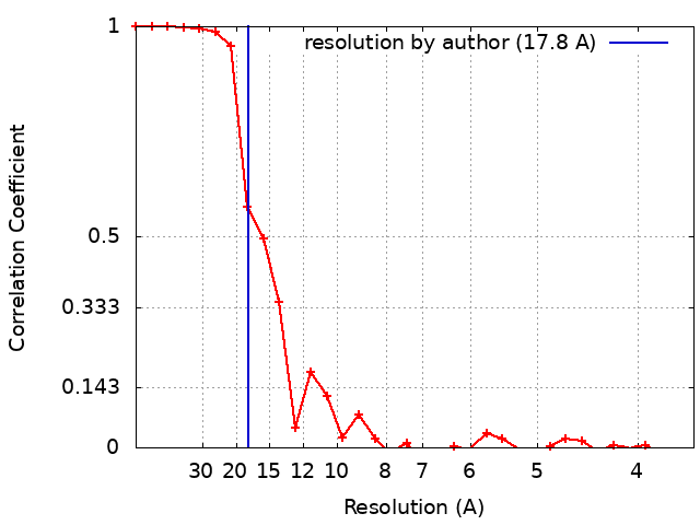
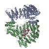
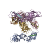
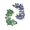
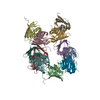
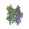
Journal: J Biol Chem / Year: 2017 Title: Single-particle electron microscopy structure of UDP-glucose:glycoprotein glucosyltransferase suggests a selectivity mechanism for misfolded proteins. Authors: Daniel Calles-Garcia / Meng Yang / Naoto Soya / Roberto Melero / Marie Ménade / Yukishige Ito / Javier Vargas / Gergely L Lukacs / Justin M Kollman / Guennadi Kozlov / Kalle Gehring / Abstract: The enzyme UDP-glucose:glycoprotein glucosyltransferase (UGGT) mediates quality control of glycoproteins in the endoplasmic reticulum by attaching glucose to -linked glycan of misfolded proteins. As ...The enzyme UDP-glucose:glycoprotein glucosyltransferase (UGGT) mediates quality control of glycoproteins in the endoplasmic reticulum by attaching glucose to -linked glycan of misfolded proteins. As a sensor, UGGT ensures that misfolded proteins are recognized by the lectin chaperones and do not leave the secretory pathway. The structure of UGGT and the mechanism of its selectivity for misfolded proteins have been unknown for 25 years. Here, we used negative-stain electron microscopy and small-angle X-ray scattering to determine the structure of UGGT from at 18-Å resolution. Three-dimensional reconstructions revealed a cage-like structure with a large central cavity. Particle classification revealed flexibility that precluded determination of a high-resolution structure. Introduction of biotinylation sites into a fungal UGGT expressed in allowed identification of the catalytic and first thioredoxin-like domains. We also used hydrogen-deuterium exchange mass spectrometry to map the binding site of an accessory protein, Sep15, to the first thioredoxin-like domain. The UGGT structural features identified suggest that the central cavity contains the catalytic site and is lined with hydrophobic surfaces. This enhances the binding of misfolded substrates with exposed hydrophobic residues and excludes folded proteins with hydrophilic surfaces. In conclusion, we have determined the UGGT structure, which enabled us to develop a plausible functional model of the mechanism for UGGT's selectivity for misfolded glycoproteins.
History
Deposition
May 3, 2017
-
Header (metadata) release
May 17, 2017
-
Map release
May 24, 2017
-
Update
Nov 25, 2020
-
Current status
Nov 25, 2020
Processing site: RCSB / Status: Released
-
Structure visualization
Movie
Surface view with section colored by density value
Details: Buffer was filtered through a 0.22um filter
Staining
Type: NEGATIVE / Material: uranyl formate Details: Freshly prepared uranyl formate at 0.75% was applied on the grids for 60s and then blotted
Grid
Model: EMS / Material: COPPER / Mesh: 400 / Support film - Material: CARBON / Support film - topology: CONTINUOUS / Support film - Film thickness: 10.0 nm / Pretreatment - Type: GLOW DISCHARGE / Pretreatment - Atmosphere: OTHER
-
Electron microscopy
Microscope
FEI TECNAI F20
Image recording
Film or detector model: GATAN ULTRASCAN 4000 (4k x 4k) / Average electron dose: 20.0 e/Å2
Electron beam
Acceleration voltage: 200 kV / Electron source: FIELD EMISSION GUN
In the structure databanks used in Yorodumi, some data are registered as the other names, "COVID-19 virus" and "2019-nCoV". Here are the details of the virus and the list of structure data.
Jan 31, 2019. EMDB accession codes are about to change! (news from PDBe EMDB page)
EMDB accession codes are about to change! (news from PDBe EMDB page)
The allocation of 4 digits for EMDB accession codes will soon come to an end. Whilst these codes will remain in use, new EMDB accession codes will include an additional digit and will expand incrementally as the available range of codes is exhausted. The current 4-digit format prefixed with “EMD-” (i.e. EMD-XXXX) will advance to a 5-digit format (i.e. EMD-XXXXX), and so on. It is currently estimated that the 4-digit codes will be depleted around Spring 2019, at which point the 5-digit format will come into force.
The EM Navigator/Yorodumi systems omit the EMD- prefix.
Related info.:Q: What is EMD? / ID/Accession-code notation in Yorodumi/EM Navigator
Yorodumi is a browser for structure data from EMDB, PDB, SASBDB, etc.
This page is also the successor to EM Navigator detail page, and also detail information page/front-end page for Omokage search.
The word "yorodu" (or yorozu) is an old Japanese word meaning "ten thousand". "mi" (miru) is to see.
Related info.:EMDB / PDB / SASBDB / Comparison of 3 databanks / Yorodumi Search / Aug 31, 2016. New EM Navigator & Yorodumi / Yorodumi Papers / Jmol/JSmol / Function and homology information / Changes in new EM Navigator and Yorodumi
 Movie
Movie Controller

 Yorodumi
 Open data
 Basic information
 Map data
 Sample
 Function and homology information
 Authors
 Canada, 1 items
  Citation


 Structure visualization
 Downloads & links
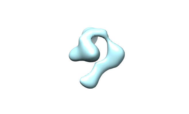 emd_8719.png
 http://ftp.pdbj.org/pub/emdb/structures/EMD-8719
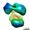


 Z (Sec.)
 Y (Row.)
 X (Col.)


 Sample components
 Spodoptera frugiperda (fall armyworm) / Recombinant cell: SF9
 Processing
 Electron microscopy
 FIELD EMISSION GUN