 Movie
Movie Controller
 Structure viewers
 About Yorodumi Papers


+Search query
-Structure paper
| Title | Single-particle electron microscopy structure of UDP-glucose:glycoprotein glucosyltransferase suggests a selectivity mechanism for misfolded proteins. |
|---|---|
| Journal, issue, pages | J Biol Chem, Vol. 292, Issue 27, Page 11499-11507, Year 2017 |
| Publish date | Jul 7, 2017 |
 Authors Authors | Daniel Calles-Garcia / Meng Yang / Naoto Soya / Roberto Melero / Marie Ménade / Yukishige Ito / Javier Vargas / Gergely L Lukacs / Justin M Kollman / Guennadi Kozlov / Kalle Gehring /     |
| PubMed Abstract | The enzyme UDP-glucose:glycoprotein glucosyltransferase (UGGT) mediates quality control of glycoproteins in the endoplasmic reticulum by attaching glucose to -linked glycan of misfolded proteins. As ...The enzyme UDP-glucose:glycoprotein glucosyltransferase (UGGT) mediates quality control of glycoproteins in the endoplasmic reticulum by attaching glucose to -linked glycan of misfolded proteins. As a sensor, UGGT ensures that misfolded proteins are recognized by the lectin chaperones and do not leave the secretory pathway. The structure of UGGT and the mechanism of its selectivity for misfolded proteins have been unknown for 25 years. Here, we used negative-stain electron microscopy and small-angle X-ray scattering to determine the structure of UGGT from at 18-Å resolution. Three-dimensional reconstructions revealed a cage-like structure with a large central cavity. Particle classification revealed flexibility that precluded determination of a high-resolution structure. Introduction of biotinylation sites into a fungal UGGT expressed in allowed identification of the catalytic and first thioredoxin-like domains. We also used hydrogen-deuterium exchange mass spectrometry to map the binding site of an accessory protein, Sep15, to the first thioredoxin-like domain. The UGGT structural features identified suggest that the central cavity contains the catalytic site and is lined with hydrophobic surfaces. This enhances the binding of misfolded substrates with exposed hydrophobic residues and excludes folded proteins with hydrophilic surfaces. In conclusion, we have determined the UGGT structure, which enabled us to develop a plausible functional model of the mechanism for UGGT's selectivity for misfolded glycoproteins. |
 External links External links | J Biol Chem / PubMed:28490633 / PubMed Central |
| Methods | EM (single particle) |
| Resolution | 17.8 - 33.0 Å |
| Structure data | 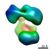 EMDB-8718: 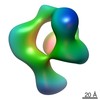 EMDB-8719: |
| Source |
|
 Penicillium chrysogenum (fungus)
