- EMDB-8660: Spacer capture and integration by a type I-F Cas1:Cas2-3 CRISPR a... -
+
Open data
ID or keywords:
Loading...
-
Basic information
Entry
Database: EMDB / ID: EMD-8660
Title
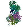
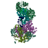
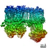
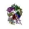
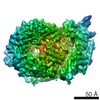
Spacer capture and integration by a type I-F Cas1:Cas2-3 CRISPR adaptation complex
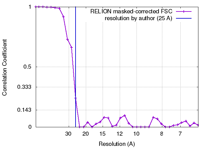
Map data
Spacer capture and integration by a type I-F Cas1:Cas2-3 CRISPR adaptation complex
Sample
Complex: type I-F Cas1:Cas2-3 CRISPR adaptation complex
Function / homology
Function and homology information
maintenance of CRISPR repeat elements / DNA endonuclease activity / defense response to virus / Hydrolases; Acting on ester bonds / hydrolase activity / DNA binding / metal ion binding Similarity search - Function
CRISPR-associated protein Cas1, YPEST subtype / CRISPR-associated endonuclease Cas1, N-terminal domain / CRISPR-associated protein Cas1 / CRISPR-associated endonuclease Cas1, C-terminal domain / CRISPR associated protein Cas1 Similarity search - Domain/homology
Journal: Proc Natl Acad Sci U S A / Year: 2017 Title: Spacer capture and integration by a type I-F Cas1-Cas2-3 CRISPR adaptation complex. Authors: Robert D Fagerlund / Max E Wilkinson / Oleg Klykov / Arjan Barendregt / F Grant Pearce / Sebastian N Kieper / Howard W R Maxwell / Angela Capolupo / Albert J R Heck / Kurt L Krause / Mihnea ...Authors: Robert D Fagerlund / Max E Wilkinson / Oleg Klykov / Arjan Barendregt / F Grant Pearce / Sebastian N Kieper / Howard W R Maxwell / Angela Capolupo / Albert J R Heck / Kurt L Krause / Mihnea Bostina / Richard A Scheltema / Raymond H J Staals / Peter C Fineran / Abstract: CRISPR-Cas adaptive immune systems capture DNA fragments from invading bacteriophages and plasmids and integrate them as spacers into bacterial CRISPR arrays. In type I-E and II-A CRISPR-Cas systems, ...CRISPR-Cas adaptive immune systems capture DNA fragments from invading bacteriophages and plasmids and integrate them as spacers into bacterial CRISPR arrays. In type I-E and II-A CRISPR-Cas systems, this adaptation process is driven by Cas1-Cas2 complexes. Type I-F systems, however, contain a unique fusion of Cas2, with the type I effector helicase and nuclease for invader destruction, Cas3. By using biochemical, structural, and biophysical methods, we present a structural model of the 400-kDa Cas1-Cas2-3 complex from with bound protospacer substrate DNA. Two Cas1 dimers assemble on a Cas2 domain dimeric core, which is flanked by two Cas3 domains forming a groove where the protospacer binds to Cas1-Cas2. We developed a sensitive in vitro assay and demonstrated that Cas1-Cas2-3 catalyzed spacer integration into CRISPR arrays. The integrase domain of Cas1 was necessary, whereas integration was independent of the helicase or nuclease activities of Cas3. Integration required at least partially duplex protospacers with free 3'-OH groups, and leader-proximal integration was stimulated by integration host factor. In a coupled capture and integration assay, Cas1-Cas2-3 processed and integrated protospacers independent of Cas3 activity. These results provide insight into the structure of protospacer-bound type I Cas1-Cas2-3 adaptation complexes and their integration mechanism.
History
Deposition
Apr 1, 2017
-
Header (metadata) release
Apr 26, 2017
-
Map release
Jun 28, 2017
-
Update
Jul 12, 2017
-
Current status
Jul 12, 2017
Processing site: RCSB / Status: Released
-
Structure visualization
Movie
Surface view with section colored by density value
In the structure databanks used in Yorodumi, some data are registered as the other names, "COVID-19 virus" and "2019-nCoV". Here are the details of the virus and the list of structure data.
Jan 31, 2019. EMDB accession codes are about to change! (news from PDBe EMDB page)
EMDB accession codes are about to change! (news from PDBe EMDB page)
The allocation of 4 digits for EMDB accession codes will soon come to an end. Whilst these codes will remain in use, new EMDB accession codes will include an additional digit and will expand incrementally as the available range of codes is exhausted. The current 4-digit format prefixed with “EMD-” (i.e. EMD-XXXX) will advance to a 5-digit format (i.e. EMD-XXXXX), and so on. It is currently estimated that the 4-digit codes will be depleted around Spring 2019, at which point the 5-digit format will come into force.
The EM Navigator/Yorodumi systems omit the EMD- prefix.
Related info.:Q: What is EMD? / ID/Accession-code notation in Yorodumi/EM Navigator
Yorodumi is a browser for structure data from EMDB, PDB, SASBDB, etc.
This page is also the successor to EM Navigator detail page, and also detail information page/front-end page for Omokage search.
The word "yorodu" (or yorozu) is an old Japanese word meaning "ten thousand". "mi" (miru) is to see.
Related info.:EMDB / PDB / SASBDB / Comparison of 3 databanks / Yorodumi Search / Aug 31, 2016. New EM Navigator & Yorodumi / Yorodumi Papers / Jmol/JSmol / Function and homology information / Changes in new EM Navigator and Yorodumi
 Movie
Movie Controller

 Yorodumi
 Open data
 Basic information
 Map data
 Sample
 Function and homology information
 Pectobacterium atrosepticum (bacteria)
 Authors
 Citation

 Structure visualization
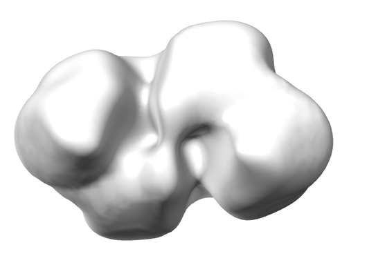
 Downloads & links
 emd_8660.png
 http://ftp.pdbj.org/pub/emdb/structures/EMD-8660


 Z (Sec.)
 Y (Row.)
 X (Col.)


 Sample components
 Processing
 Electron microscopy
 FIELD EMISSION GUN