 Movie
Movie Controller


+ Open data

- Basic information
| Entry | Database: EMDB / ID: EMD-8297 | |||||||||
|---|---|---|---|---|---|---|---|---|---|---|
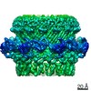
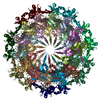
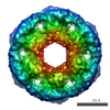
| Title | P. aeruginosa Type IVa secretin PilQ | |||||||||
 Map data Map data | PilQ oligomer with C7 symmetry | |||||||||
 Sample Sample |
| |||||||||
| Biological species |   Pseudomonas aeruginosa (bacteria) Pseudomonas aeruginosa (bacteria) | |||||||||
| Method | single particle reconstruction / cryo EM / Resolution: 7.4 Å | |||||||||
 Authors Authors | Koo J / Rubinstein JL / Burrows LL / Howell PL | |||||||||
 Citation Citation | Journal: Structure / Year: 2016 Title: Structure of the Pseudomonas aeruginosa Type IVa Pilus Secretin at 7.4 Å. Authors: Jason Koo / Ryan P Lamers / John L Rubinstein / Lori L Burrows / P Lynne Howell /  Abstract: Type IVa pili (T4aP) function as bacterial virulence factors. T4aP pass through the outer membranes of Gram-negative bacteria via homo-oligomeric secretins. We present a 7.4 Å cryoelectron ...Type IVa pili (T4aP) function as bacterial virulence factors. T4aP pass through the outer membranes of Gram-negative bacteria via homo-oligomeric secretins. We present a 7.4 Å cryoelectron microscopy structure of the Pseudomonas aeruginosa PilQ secretin. Peripheral and internal features show that the secretin is composed of 14 subunits with C7 symmetry. The channel is a ribbed cylinder with central peripheral spokes and a central gate closed on the periplasmic side. The structure suggests that during pilus extrusion, the central gate is displaced to the interior walls and that no additional conformational changes are required, as the internal diameter can accommodate the pilus. The N1 domain was resolved, while the N0 and the N-terminal β-domains proposed to bind peptidoglycan were absent in class average images and the final 3D map, indicating a high flexibility. These data provide the highest-resolution structure to date of a T4aP secretin. | |||||||||
| History |
|
- Structure visualization
| Movie |
 Movie viewer Movie viewer |
|---|---|
| Structure viewer | EM map: SurfViewMolmilJmol/JSmol |
| Supplemental images |

- Downloads & links
-EMDB archive
| Map data | emd_8297.map.gz | 28.5 MB | EMDB map data format | |
|---|---|---|---|---|
| Header (meta data) | emd-8297-v30.xmlemd-8297.xml | 12.4 KB 12.4 KB | Display Display | EMDB header |
| Images | 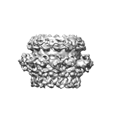 emd_8297.png emd_8297.png | 38.4 KB | ||
| Archive directory |  http://ftp.pdbj.org/pub/emdb/structures/EMD-8297ftp://ftp.pdbj.org/pub/emdb/structures/EMD-8297 http://ftp.pdbj.org/pub/emdb/structures/EMD-8297ftp://ftp.pdbj.org/pub/emdb/structures/EMD-8297 | HTTPS FTP |
-Related structure data
| Similar structure data |
|---|


-Links
| EMDB pages | EMDB (EBI/PDBe) / EMDataResource |
|---|
-Map
| File | Download / File: emd_8297.map.gz / Format: CCP4 / Size: 30.5 MB / Type: IMAGE STORED AS FLOATING POINT NUMBER (4 BYTES) | ||||||||||||||||||||||||||||||||||||||||||||||||||||||||||||||||||||
|---|---|---|---|---|---|---|---|---|---|---|---|---|---|---|---|---|---|---|---|---|---|---|---|---|---|---|---|---|---|---|---|---|---|---|---|---|---|---|---|---|---|---|---|---|---|---|---|---|---|---|---|---|---|---|---|---|---|---|---|---|---|---|---|---|---|---|---|---|---|
| Annotation | PilQ oligomer with C7 symmetry | ||||||||||||||||||||||||||||||||||||||||||||||||||||||||||||||||||||
| Projections & slices | Image control
Images are generated by Spider. | ||||||||||||||||||||||||||||||||||||||||||||||||||||||||||||||||||||
| Voxel size | X=Y=Z: 1.45 Å | ||||||||||||||||||||||||||||||||||||||||||||||||||||||||||||||||||||
| Density |
| ||||||||||||||||||||||||||||||||||||||||||||||||||||||||||||||||||||
| Symmetry | Space group: 1 | ||||||||||||||||||||||||||||||||||||||||||||||||||||||||||||||||||||
| Details | EMDB XML:
CCP4 map header:
| ||||||||||||||||||||||||||||||||||||||||||||||||||||||||||||||||||||
 Z (Sec.)
 Y (Row.)
 X (Col.)


-Supplemental data
- Sample components
-Entire : Pseudomonas aeruginosa Type IVa pilus secretin, PilQ
| Entire | Name: Pseudomonas aeruginosa Type IVa pilus secretin, PilQ |
|---|---|
| Components |
|
-Supramolecule #1: Pseudomonas aeruginosa Type IVa pilus secretin, PilQ
| Supramolecule | Name: Pseudomonas aeruginosa Type IVa pilus secretin, PilQ / type: complex / ID: 1 / Parent: 0 |
|---|---|
| Source (natural) | Organism: Pseudomonas aeruginosa (bacteria) |
| Recombinant expression | Organism: Pseudomonas aeruginosa (bacteria) / Recombinant plasmid: pUCP20Gm-pilQ2xHis8 |
-Experimental details
-Structure determination
| Method | cryo EM |
|---|---|
 Processing Processing | single particle reconstruction |
| Aggregation state | particle |
-Sample preparation
| Concentration | 0.03 mg/mL | ||||||||||||
|---|---|---|---|---|---|---|---|---|---|---|---|---|---|
| Buffer | pH: 8 Component:
| ||||||||||||
| Grid | Model: Maxtaform / Material: COPPER/RHODIUM / Mesh: 400 / Support film - Material: CARBON / Support film - topology: CONTINUOUS / Support film - Film thickness: 7.5 nm / Pretreatment - Type: GLOW DISCHARGE / Pretreatment - Atmosphere: AIR / Pretreatment - Pressure: 0.3 kPa | ||||||||||||
| Vitrification | Cryogen name: ETHANE-PROPANE / Chamber humidity: 95 % / Chamber temperature: 277 K / Instrument: FEI VITROBOT MARK III Details: Vitrobot modified to use ethane-propane mixture at liquid nitrogen temperature. |
- Electron microscopy
| Microscope | FEI TECNAI F20 |
|---|---|
| Image recording | Film or detector model: GATAN K2 SUMMIT (4k x 4k) / Detector mode: COUNTING / Digitization - Sampling interval: 5.0 µm / Digitization - Frames/image: 1-30 / Number real images: 2419 / Average exposure time: 15.0 sec. / Average electron dose: 35.6 e/Å2 |
| Electron beam | Acceleration voltage: 200 kV / Electron source:  FIELD EMISSION GUN FIELD EMISSION GUN |
| Electron optics | C2 aperture diameter: 30.0 µm / Calibrated defocus max: 4.6 µm / Calibrated defocus min: 0.7 µm / Calibrated magnification: 34483 / Illumination mode: FLOOD BEAM / Imaging mode: BRIGHT FIELD / Cs: 2.0 mm / Nominal magnification: 25000 |
| Sample stage | Specimen holder model: GATAN 626 SINGLE TILT LIQUID NITROGEN CRYO TRANSFER HOLDER Cooling holder cryogen: NITROGEN |
| Experimental equipment |  Model: Tecnai F20 / Image courtesy: FEI Company |
