 ムービー
ムービー コントローラー

[日本語] English
 万見
万見- EMDB-8278: Structure of Nanoparticle Released from Enveloped Protein Nanoparticle -
+ データを開く

- 基本情報
基本情報
| 登録情報 | データベース: EMDB / ID: EMD-8278 | ||||||||||||||||||
|---|---|---|---|---|---|---|---|---|---|---|---|---|---|---|---|---|---|---|---|
| タイトル | Structure of Nanoparticle Released from Enveloped Protein Nanoparticle | ||||||||||||||||||
 マップデータ マップデータ | Icosahedrally-symmetrized, average reconstruction of nanoparticle released from EPN | ||||||||||||||||||
 試料 試料 |
| ||||||||||||||||||
 キーワード キーワード | protein design / icosahedral assemblies / cell transduction / enveloped viruses / virus assembly / enveloped protein / nanoparticle / STRUCTURAL PROTEIN | ||||||||||||||||||
| 機能・相同性 |  機能・相同性情報 機能・相同性情報D-galactonate catabolic process / 2-dehydro-3-deoxy-6-phosphogalactonate aldolase activity / viral budding via host ESCRT complex / host multivesicular body / viral nucleocapsid / viral translational frameshifting / host cell nucleus / host cell plasma membrane / virion membrane / structural molecule activity ...D-galactonate catabolic process / 2-dehydro-3-deoxy-6-phosphogalactonate aldolase activity / viral budding via host ESCRT complex / host multivesicular body / viral nucleocapsid / viral translational frameshifting / host cell nucleus / host cell plasma membrane / virion membrane / structural molecule activity / RNA binding / zinc ion binding / identical protein binding / membrane 類似検索 - 分子機能 | ||||||||||||||||||
| 生物種 |   Thermotoga maritima (バクテリア) / Thermotoga maritima (バクテリア) /  Human immunodeficiency virus type 1 group M subtype B (isolate BH10) (ヒト免疫不全ウイルス) Human immunodeficiency virus type 1 group M subtype B (isolate BH10) (ヒト免疫不全ウイルス) | ||||||||||||||||||
| 手法 | 単粒子再構成法 / クライオ電子顕微鏡法 / 解像度: 5.7 Å | ||||||||||||||||||
 データ登録者 データ登録者 | Votteler J / Ogohara C | ||||||||||||||||||
| 資金援助 |  ドイツ, ドイツ,  米国, 5件 米国, 5件
| ||||||||||||||||||
 引用 引用 | ジャーナル: Nature / 年: 2016 タイトル: Designed proteins induce the formation of nanocage-containing extracellular vesicles. 著者: Jörg Votteler / Cassandra Ogohara / Sue Yi / Yang Hsia / Una Nattermann / David M Belnap / Neil P King / Wesley I Sundquist / 要旨: Complex biological processes are often performed by self-organizing nanostructures comprising multiple classes of macromolecules, such as ribosomes (proteins and RNA) or enveloped viruses (proteins, ...Complex biological processes are often performed by self-organizing nanostructures comprising multiple classes of macromolecules, such as ribosomes (proteins and RNA) or enveloped viruses (proteins, nucleic acids and lipids). Approaches have been developed for designing self-assembling structures consisting of either nucleic acids or proteins, but strategies for engineering hybrid biological materials are only beginning to emerge. Here we describe the design of self-assembling protein nanocages that direct their own release from human cells inside small vesicles in a manner that resembles some viruses. We refer to these hybrid biomaterials as 'enveloped protein nanocages' (EPNs). Robust EPN biogenesis requires protein sequence elements that encode three distinct functions: membrane binding, self-assembly, and recruitment of the endosomal sorting complexes required for transport (ESCRT) machinery. A variety of synthetic proteins with these functional elements induce EPN biogenesis, highlighting the modularity and generality of the design strategy. Biochemical analyses and cryo-electron microscopy reveal that one design, EPN-01, comprises small (~100 nm) vesicles containing multiple protein nanocages that closely match the structure of the designed 60-subunit self-assembling scaffold. EPNs that incorporate the vesicular stomatitis viral glycoprotein can fuse with target cells and deliver their contents, thereby transferring cargoes from one cell to another. These results show how proteins can be programmed to direct the formation of hybrid biological materials that perform complex tasks, and establish EPNs as a class of designed, modular, genetically-encoded nanomaterials that can transfer molecules between cells. | ||||||||||||||||||
| 履歴 |
|
- 構造の表示
構造の表示
| ムービー |
ムービービューア |
|---|---|
| 構造ビューア | EMマップ: SurfViewMolmilJmol/JSmol |
| 添付画像 |

- ダウンロードとリンク
-EMDBアーカイブ
| マップデータ | emd_8278.map.gz | 28.3 MB | EMDBマップデータ形式 | |
|---|---|---|---|---|
| ヘッダ (付随情報) | emd-8278-v30.xmlemd-8278.xml | 14.8 KB 14.8 KB | 表示 表示 | EMDBヘッダ |
| 画像 | 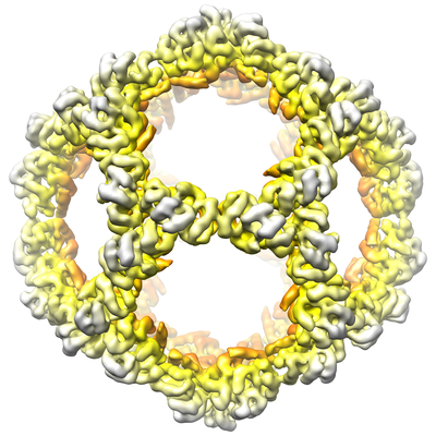 emd_8278.png emd_8278.png | 196.4 KB | ||
| Filedesc metadata | emd-8278.cif.gz | 6.1 KB | ||
| アーカイブディレクトリ |  http://ftp.pdbj.org/pub/emdb/structures/EMD-8278ftp://ftp.pdbj.org/pub/emdb/structures/EMD-8278 http://ftp.pdbj.org/pub/emdb/structures/EMD-8278ftp://ftp.pdbj.org/pub/emdb/structures/EMD-8278 | HTTPS FTP |
-検証レポート
| 文書・要旨 | emd_8278_validation.pdf.gz | 498.2 KB | 表示 | EMDB検証レポート |
|---|---|---|---|---|
| 文書・詳細版 | emd_8278_full_validation.pdf.gz | 497.8 KB | 表示 | |
| XML形式データ | emd_8278_validation.xml.gz | 6.5 KB | 表示 | |
| CIF形式データ | emd_8278_validation.cif.gz | 7.5 KB | 表示 | |
| アーカイブディレクトリ | https://ftp.pdbj.org/pub/emdb/validation_reports/EMD-8278ftp://ftp.pdbj.org/pub/emdb/validation_reports/EMD-8278 | HTTPS FTP |
-関連構造データ

-リンク
| EMDBのページ | EMDB (EBI/PDBe) / EMDataResource |
|---|---|
| 「今月の分子」の関連する項目 |


-マップ
| ファイル | ダウンロード / ファイル: emd_8278.map.gz / 形式: CCP4 / 大きさ: 103 MB / タイプ: IMAGE STORED AS FLOATING POINT NUMBER (4 BYTES) | ||||||||||||||||||||||||||||||||||||||||||||||||||||||||||||||||||||
|---|---|---|---|---|---|---|---|---|---|---|---|---|---|---|---|---|---|---|---|---|---|---|---|---|---|---|---|---|---|---|---|---|---|---|---|---|---|---|---|---|---|---|---|---|---|---|---|---|---|---|---|---|---|---|---|---|---|---|---|---|---|---|---|---|---|---|---|---|---|
| 注釈 | Icosahedrally-symmetrized, average reconstruction of nanoparticle released from EPN | ||||||||||||||||||||||||||||||||||||||||||||||||||||||||||||||||||||
| 投影像・断面図 | 画像のコントロール
画像は Spider により作成 | ||||||||||||||||||||||||||||||||||||||||||||||||||||||||||||||||||||
| ボクセルのサイズ | X=Y=Z: 1.193 Å | ||||||||||||||||||||||||||||||||||||||||||||||||||||||||||||||||||||
| 密度 |
| ||||||||||||||||||||||||||||||||||||||||||||||||||||||||||||||||||||
| 対称性 | 空間群: 1 | ||||||||||||||||||||||||||||||||||||||||||||||||||||||||||||||||||||
| 詳細 | EMDB XML:
CCP4マップ ヘッダ情報:
| ||||||||||||||||||||||||||||||||||||||||||||||||||||||||||||||||||||
 Z (Sec.)
 Y (Row.)
 X (Col.)


-添付データ
- 試料の構成要素
-全体 : EPN-01*
| 全体 | 名称: EPN-01* |
|---|---|
| 要素 |
|
-超分子 #1: EPN-01*
| 超分子 | 名称: EPN-01* / タイプ: complex / ID: 1 / 親要素: 0 / 含まれる分子: all |
|---|---|
| 由来(天然) | 生物種: Thermotoga maritima (バクテリア) |
-分子 #1: EPN-01*
| 分子 | 名称: EPN-01* / タイプ: protein_or_peptide / ID: 1 / コピー数: 1 / 光学異性体: LEVO |
|---|---|
| 由来(天然) | 生物種: Human immunodeficiency virus type 1 group M subtype B (isolate BH10) (ヒト免疫不全ウイルス) 株: isolate BH10 |
| 分子量 | 理論値: 30.30492 KDa |
| 組換発現 | 生物種:  Homo sapiens (ヒト) Homo sapiens (ヒト) |
| 配列 | 文字列: MGARASGSKS GSGSDSGSKM EELFKKHKIV AVLRANSVEE AKKKALAVFL GGVHLIEITF TVPDADTVIK ELSFLKEMGA IIGAGTVTS VEQCRKAVES GAEFIVSPHL DEEISQFCKE KGVFYMPGVM TPTELVKAMK LGHTILKLFP GEVVGPQFVK A MKGPFPNV ...文字列: MGARASGSKS GSGSDSGSKM EELFKKHKIV AVLRANSVEE AKKKALAVFL GGVHLIEITF TVPDADTVIK ELSFLKEMGA IIGAGTVTS VEQCRKAVES GAEFIVSPHL DEEISQFCKE KGVFYMPGVM TPTELVKAMK LGHTILKLFP GEVVGPQFVK A MKGPFPNV KFVPTGGVNL DNVCEWFKAG VLAVGVGSAL VKGTPVEVAE KAKAFVEKIR GCTEQKLISE EDLQSRPEPT AP PEESFRS GVETTTPPQK QEPIDKELYP LTSLRSLFGN DPSSQ UniProtKB: 2-dehydro-3-deoxyphosphogluconate aldolase/4-hydroxy-2-oxoglutarate aldolase, Gag polyprotein |
-実験情報
-構造解析
| 手法 | クライオ電子顕微鏡法 |
|---|---|
 解析 解析 | 単粒子再構成法 |
| 試料の集合状態 | particle |
-試料調製
| 濃度 | 0.2 mg/mL |
|---|---|
| 緩衝液 | pH: 7.5 / 詳細: Phosphate-buffered saline |
| グリッド | モデル: Quantifoil R2/2 / 材質: COPPER / 支持フィルム - 材質: CARBON / 支持フィルム - トポロジー: HOLEY ARRAY / 前処理 - タイプ: GLOW DISCHARGE |
| 凍結 | 凍結剤: ETHANE / チャンバー内湿度: 80 % / チャンバー内温度: 277 K / 装置: FEI VITROBOT MARK II / 詳細: 11 second blot, 0 mm offset. |
| 詳細 | EPN nanoparticles released from vesicles by detergent treatment (0.75% CHAPS) |
- 電子顕微鏡法
| 顕微鏡 | FEI TECNAI F20 |
|---|---|
| 撮影 | フィルム・検出器のモデル: GATAN K2 SUMMIT (4k x 4k) 検出モード: SUPER-RESOLUTION / デジタル化 - サイズ - 横: 7420 pixel / デジタル化 - サイズ - 縦: 7676 pixel / 平均電子線量: 2.0 e/Å2 / 詳細: Electron dose at specimen was not recorded. |
| 電子線 | 加速電圧: 200 kV / 電子線源:  FIELD EMISSION GUN FIELD EMISSION GUN |
| 電子光学系 | 最大 デフォーカス(補正後): 3.3000000000000003 µm 最小 デフォーカス(補正後): 0.7000000000000001 µm 倍率(補正後): 41911 / 照射モード: FLOOD BEAM / 撮影モード: BRIGHT FIELD / Cs: 2.0 mm |
| 試料ステージ | 試料ホルダーモデル: GATAN 626 SINGLE TILT LIQUID NITROGEN CRYO TRANSFER HOLDER ホルダー冷却材: NITROGEN |
| 実験機器 |  モデル: Tecnai F20 / 画像提供: FEI Company |
+画像解析
-原子モデル構築 1
| 精密化 | プロトコル: RIGID BODY FIT |
|---|---|
| 得られたモデル | 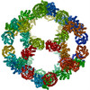 PDB-5kp9: |
