 Movie
Movie Controller


+ Open data

- Basic information
| Entry |  | |||||||||
|---|---|---|---|---|---|---|---|---|---|---|
| Title | tail fiber of F2-pyocin | |||||||||
 Map data Map data | ||||||||||
 Sample Sample |
| |||||||||
 Keywords Keywords | pyocin / phage / cap / VIRAL PROTEIN | |||||||||
| Function / homology | : / :  Function and homology information Function and homology information | |||||||||
| Biological species |  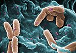 Pseudomonas aeruginosa (bacteria) Pseudomonas aeruginosa (bacteria) | |||||||||
| Method | single particle reconstruction / cryo EM / Resolution: 3.26 Å | |||||||||
 Authors Authors | Gu ZW / Xie YF / Wang JW | |||||||||
| Funding support |  China, 1 items China, 1 items
| |||||||||
 Citation Citation | Journal: Adv Sci (Weinh) / Year: 2026 Title: F-Type Pyocin Versus Phage λ Tail: Conserved Hub, Divergent Fibers. Authors: Zhiwei Gu / Yufan Xie / Lanxin Wang / Luyan Ma / Xiaofei Ge / Jiawei Wang / Abstract: Bacteriocins are ribosomally synthesized antimicrobial peptides or proteins that offer an alternative to conventional antibiotics against multidrug-resistant pathogens. Phage tail-like bacteriocins ...Bacteriocins are ribosomally synthesized antimicrobial peptides or proteins that offer an alternative to conventional antibiotics against multidrug-resistant pathogens. Phage tail-like bacteriocins (tailocins) are classified into rigid R-type and flexible F-type variants. While R-type pyocins from Pseudomonas aeruginosa are well-characterized, F-type pyocins remain poorly understood, especially with respect to the molecular mechanisms for their Gram-negative bactericidal activity. Here, we report cryo-electron microscopy structures of the F-type pyocin from P. aeruginosa ATCC 15442 at 2.29-3.26 Å resolution, encompassing three modular components: the tail cap, tail tip, and tail fiber. Structural comparisons with bacteriophage λ reveal a conserved tail tip architecture, including the baseplate hub proteins, distal tail protein, tail assembly protein, and tape measure protein. Unexpectedly, we identify three trimeric side fibers that attach not to the distal tail protein, as in canonical systems, but to the α-helical shaft of the central fiber, indicating a previously unrecognized attachment mode. The receptor-binding domain of the side fiber shares structural similarity with LPS-recognizing domains of R-type pyocins. Together, these results define the structural basis of F-type pyocin assembly and host recognition, reveal conserved and unique features relative to phage λ, and provide a framework for engineering tailocins as precision antimicrobials against drug-resistant P. aeruginosa. | |||||||||
| History |
|
- Structure visualization
| Supplemental images |
|---|

- Downloads & links
-EMDB archive
| Map data | emd_80404.map.gz | 483.9 MB | EMDB map data format | |
|---|---|---|---|---|
| Header (meta data) | emd-80404-v30.xmlemd-80404.xml | 19.7 KB 19.7 KB | Display Display | EMDB header |
| FSC (resolution estimation) | emd_80404_fsc.xml | 16.9 KB | Display | FSC data file |
| Images |  emd_80404.png emd_80404.png | 48.2 KB | ||
| Filedesc metadata | emd-80404.cif.gz | 6.6 KB | ||
| Others | emd_80404_half_map_1.map.gzemd_80404_half_map_2.map.gz | 474.6 MB 474.6 MB | ||
| Archive directory |  https://data.pdbj.org/pub/emdb/structures/EMD-80404ftp://data.pdbj.org/pub/emdb/structures/EMD-80404 https://data.pdbj.org/pub/emdb/structures/EMD-80404ftp://data.pdbj.org/pub/emdb/structures/EMD-80404 | HTTPS FTP |
-Related structure data
| Related structure data |  25vjMC  25vdC  25veC  25vfC  26ciC M: atomic model generated by this map C: citing same article ( |
|---|---|
| Similar structure data |


-Links
| EMDB pages | EMDB (EBI/PDBe) / EMDataResource |
|---|
-Map
| File | Download / File: emd_80404.map.gz / Format: CCP4 / Size: 512 MB / Type: IMAGE STORED AS FLOATING POINT NUMBER (4 BYTES) | ||||||||||||||||||||||||||||||||||||
|---|---|---|---|---|---|---|---|---|---|---|---|---|---|---|---|---|---|---|---|---|---|---|---|---|---|---|---|---|---|---|---|---|---|---|---|---|---|
| Projections & slices | Image control
Images are generated by Spider. | ||||||||||||||||||||||||||||||||||||
| Voxel size | X=Y=Z: 0.926 Å | ||||||||||||||||||||||||||||||||||||
| Density |
| ||||||||||||||||||||||||||||||||||||
| Symmetry | Space group: 1 | ||||||||||||||||||||||||||||||||||||
| Details | EMDB XML:
|
 Z (Sec.)
 Y (Row.)
 X (Col.)


-Supplemental data
-Half map: #1
| File | emd_80404_half_map_1.map | ||||||||||||
|---|---|---|---|---|---|---|---|---|---|---|---|---|---|
| Projections & Slices |
| ||||||||||||
| Density Histograms |


-Half map: #2
| File | emd_80404_half_map_2.map | ||||||||||||
|---|---|---|---|---|---|---|---|---|---|---|---|---|---|
| Projections & Slices |
| ||||||||||||
| Density Histograms |


- Sample components
-Entire : F2-pyocin Cap
| Entire | Name: F2-pyocin Cap |
|---|---|
| Components |
|
-Supramolecule #1: F2-pyocin Cap
| Supramolecule | Name: F2-pyocin Cap / type: complex / ID: 1 / Parent: 0 / Macromolecule list: all |
|---|---|
| Source (natural) | Organism: Pseudomonas aeruginosa (bacteria) |
-Macromolecule #1: tail fiber protein of F2-pyocin
| Macromolecule | Name: tail fiber protein of F2-pyocin / type: protein_or_peptide / ID: 1 / Number of copies: 3 / Enantiomer: LEVO |
|---|---|
| Source (natural) | Organism: Pseudomonas aeruginosa (bacteria) |
| Molecular weight | Theoretical: 130.815414 KDa |
| Recombinant expression | Organism: Pseudomonas aeruginosa (bacteria) |
| Sequence | String: MNKTITGHKG GSKKPRQPVE MPDSVRSIAR AKILLALGEG EFDGGVDGRS IYLDDTPLLA ADGSVNFPGV TWEFRPGSVD QEHIAGVPA VENELAVGVE LKSDAPWVRA VSNTQLSAVR LRLSWPAIQR QQENGDVVGY RIDYAIDIAV DGGAWQEALK A SLDDKSTS ...String: MNKTITGHKG GSKKPRQPVE MPDSVRSIAR AKILLALGEG EFDGGVDGRS IYLDDTPLLA ADGSVNFPGV TWEFRPGSVD QEHIAGVPA VENELAVGVE LKSDAPWVRA VSNTQLSAVR LRLSWPAIQR QQENGDVVGY RIDYAIDIAV DGGAWQEALK A SLDDKSTS RYERSHRVDL PEARSGWQVR VRRLTPNQNN NRVADTMRVE AITEVIDAKL RYPNTALLFV EFDASQFQSI PQ ISVEARG RRVRVPSNYD PQTRSYSGTW DGSFKSAWTS NPAWHWYDIV LHKRFGLGRR IDASMVDKWS LYRIAQYCDQ SVP DGKGGQ EPRFSCNLYL QSRAEAWTVL RDLAAIFRGM SYWSGAEMVA VSDMPEDEAY TFSPSNTVRG DDGSHFNYSS SRQR DRHTL ALVNYDNPGN GYQSQPVAVN NDRAQRRYGI SQLEITAIGC TSEGEAQRRG QWALLTEELE QDAVTFRTGM DGRGL APGK IIAVADPVKS GKQIGGRLSA VDGRALTLDR DVEARPGDRL LVNLPNGKAE ARSVQSVVGR VLSVTASYSE TPRPQG QWA LQSNSLTTQR FRIMSITRPE DNLFEITALQ HNASKFDAID NGARIELPPV TSIPPGVQAP PQNVRIKAFT KVDQGLA VT SLSASWDAAP NAVAYEAEWR KDSGNWVRVP RTSALGFDVP GIYAGRYLVR VRALNVMEVG SVYASSEETA LEGKTTPP P ALAYLRCVAG PWRIGLEWGF PASGAADTAY TEIQQSATPG GSEETARALG LFAYPGNTHL VSPIPAGERL AFRGRLIDR SGNVGAWSNW VTGTSSSDAS EYNQLITQEY VESALGQQFF SDIERMQVDI GGLQKQVGDL ADVLLYDPAK VYAKNDMVRQ GQRLYQALK AVPAKTAPPN AAYWSDIGQS LETANGLAQQ VASHTAEISE LDGSLTAQAS RLGVLLAATR DDADDGNGAM A DALRGWKT VARAAQEETV RATENEAQAT RTTLLEARTA DAEGRIATVE RVATSDRQAT AQRLDQLSAS IGGTAASLQS EQ TARANAD SALAQRIDTV QASTDTNSAA IQTTSQAVTS LDGNVKAMYS VKLQAHANGQ KYAAGWQLGF DSGTSVSTMA FQA DRFIWF DSSSGTAVAP VSIVNGQMFI KNALIQDGAI DNAKIGNVIQ SNAMSNNGMP IWRLDKAGTY TVRDSAGQVR VEMG LLQS UniProtKB: UNIPROTKB: A0A5E5R453 |
-Macromolecule #2: F2-pyocin side fiber
| Macromolecule | Name: F2-pyocin side fiber / type: protein_or_peptide / ID: 2 / Number of copies: 9 / Enantiomer: LEVO |
|---|---|
| Source (natural) | Organism: Pseudomonas aeruginosa (bacteria) |
| Molecular weight | Theoretical: 38.697773 KDa |
| Recombinant expression | Organism: Pseudomonas aeruginosa (bacteria) |
| Sequence | String: MAWHSKGSVS VTLNSEAVLG NATDFIANVR TGDAFRGPDG RWYEITNVTS ATVISIKPNY QGATASGQVY AVVPIHGYSK NLADQFRDI NNQWGATLAG IKPWAVSSTG QQAQADMGIS AVGRALNNAS TPANALSYLG GVAPNQMGWA GNAMNTADLD S LTVSGLYA ...String: MAWHSKGSVS VTLNSEAVLG NATDFIANVR TGDAFRGPDG RWYEITNVTS ATVISIKPNY QGATASGQVY AVVPIHGYSK NLADQFRDI NNQWGATLAG IKPWAVSSTG QQAQADMGIS AVGRALNNAS TPANALSYLG GVAPNQMGWA GNAMNTADLD S LTVSGLYA HGTAVPSPVN NAQGYVLHMQ HGNPDFAVQQ WYQLNSATGQ YMRIKAGGNW SRWVLQYSQF NLVGLASFDA SN NPSGAII QRGGTVGFNE YVRYADGTQI CWGNTTTNVG ATMAYQPAGT LSFYITPVAY SWGFPVSFSR PPSVMVNPMR AAG NNASRP WGSTMSVTET LFSWYGYDTA SVASGMAASY VAMGRWN UniProtKB: UNIPROTKB: A0A2V3GUQ2 |
-Experimental details
-Structure determination
| Method | cryo EM |
|---|---|
 Processing Processing | single particle reconstruction |
| Aggregation state | particle |
-Sample preparation
| Buffer | pH: 7.5 |
|---|---|
| Vitrification | Cryogen name: ETHANE |
- Electron microscopy
| Microscope | FEI POLARA 300 |
|---|---|
| Image recording | Film or detector model: FEI FALCON IV (4k x 4k) / Average electron dose: 49.91 e/Å2 |
| Electron beam | Acceleration voltage: 300 kV / Electron source:  FIELD EMISSION GUN FIELD EMISSION GUN |
| Electron optics | Illumination mode: FLOOD BEAM / Imaging mode: BRIGHT FIELD / Nominal defocus max: 2.0 µm / Nominal defocus min: 1.5 µm |
| Experimental equipment | 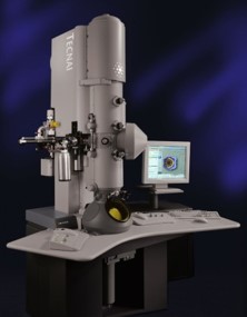 Model: Tecnai Polara / Image courtesy: FEI Company |

