 Movie
Movie Controller

[English] 日本語
 Yorodumi
- EMDB-7947: Classification of Single Particles from Human Cell Extract Reveal... -
+ Open data

- Basic information
| Entry | Database: EMDB / ID: EMD-7947 | |||||||||
|---|---|---|---|---|---|---|---|---|---|---|
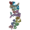
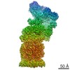
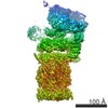
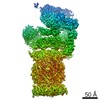
| Title | Classification of Single Particles from Human Cell Extract Reveals Distinct Structures | |||||||||
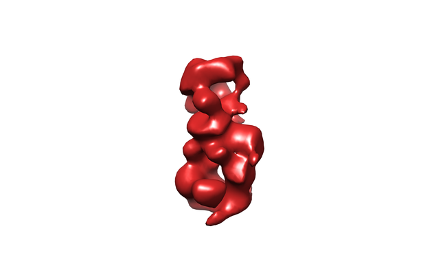
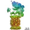
 Map data Map data | 3D class of 26S Proteasome | |||||||||
 Sample Sample |
| |||||||||
| Biological species |  Homo sapiens (human) Homo sapiens (human) | |||||||||
| Method | single particle reconstruction / negative staining / Resolution: 31.0 Å | |||||||||
 Authors Authors | Verbeke EJ / Mallam AL / Drew K / Marcotte EM / Taylor DW | |||||||||
 Citation Citation | Journal: Cell Rep / Year: 2018 Title: Classification of Single Particles from Human Cell Extract Reveals Distinct Structures. Authors: Eric J Verbeke / Anna L Mallam / Kevin Drew / Edward M Marcotte / David W Taylor /  Abstract: Multi-protein complexes are necessary for nearly all cellular processes, and understanding their structure is required for elucidating their function. Current high-resolution strategies in structural ...Multi-protein complexes are necessary for nearly all cellular processes, and understanding their structure is required for elucidating their function. Current high-resolution strategies in structural biology are effective but lag behind other fields (e.g., genomics and proteomics) due to their reliance on purified samples rather than heterogeneous mixtures. Here, we present a method combining single-particle analysis by electron microscopy with protein identification by mass spectrometry to structurally characterize macromolecular complexes from human cell extract. We identify HSP60 through two-dimensional classification and obtain three-dimensional structures of native proteasomes directly from ab initio classification of a heterogeneous mixture of protein complexes. In addition, we reveal an ∼1-MDa-size structure of unknown composition and reference our proteomics data to suggest possible identities. Our study shows the power of using a shotgun approach to electron microscopy (shotgun EM) when coupled with mass spectrometry as a tool to uncover the structures of macromolecular machines. | |||||||||
| History |
|
- Structure visualization
| Movie |
 Movie viewer Movie viewer |
|---|---|
| Structure viewer | EM map: SurfViewMolmilJmol/JSmol |
| Supplemental images |
- Downloads & links
-EMDB archive
| Map data | emd_7947.map.gz | 3.5 MB | EMDB map data format | |
|---|---|---|---|---|
| Header (meta data) | emd-7947-v30.xmlemd-7947.xml | 9 KB 9 KB | Display Display | EMDB header |
| Images |  emd_7947.png emd_7947.png | 40.9 KB | ||
| Archive directory |  http://ftp.pdbj.org/pub/emdb/structures/EMD-7947ftp://ftp.pdbj.org/pub/emdb/structures/EMD-7947 http://ftp.pdbj.org/pub/emdb/structures/EMD-7947ftp://ftp.pdbj.org/pub/emdb/structures/EMD-7947 | HTTPS FTP |
-Related structure data

-Links
| EMDB pages | EMDB (EBI/PDBe) / EMDataResource |
|---|
-Map
| File | Download / File: emd_7947.map.gz / Format: CCP4 / Size: 11.4 MB / Type: IMAGE STORED AS FLOATING POINT NUMBER (4 BYTES) | ||||||||||||||||||||||||||||||||||||||||||||||||||||||||||||
|---|---|---|---|---|---|---|---|---|---|---|---|---|---|---|---|---|---|---|---|---|---|---|---|---|---|---|---|---|---|---|---|---|---|---|---|---|---|---|---|---|---|---|---|---|---|---|---|---|---|---|---|---|---|---|---|---|---|---|---|---|---|
| Annotation | 3D class of 26S Proteasome | ||||||||||||||||||||||||||||||||||||||||||||||||||||||||||||
| Projections & slices | Image control
Images are generated by Spider. | ||||||||||||||||||||||||||||||||||||||||||||||||||||||||||||
| Voxel size | X=Y=Z: 3.6 Å | ||||||||||||||||||||||||||||||||||||||||||||||||||||||||||||
| Density |
| ||||||||||||||||||||||||||||||||||||||||||||||||||||||||||||
| Symmetry | Space group: 1 | ||||||||||||||||||||||||||||||||||||||||||||||||||||||||||||
| Details | EMDB XML:
CCP4 map header:
| ||||||||||||||||||||||||||||||||||||||||||||||||||||||||||||
 Z (Sec.)
 Y (Row.)
 X (Col.)


-Supplemental data
- Sample components
-Entire : 26S Proteasome
| Entire | Name: 26S Proteasome |
|---|---|
| Components |
|
-Supramolecule #1: 26S Proteasome
| Supramolecule | Name: 26S Proteasome / type: complex / ID: 1 / Parent: 0 |
|---|---|
| Source (natural) | Organism: Homo sapiens (human) / Strain: HEK293T |
| Molecular weight | Theoretical: 2.5 MDa |
-Experimental details
-Structure determination
| Method | negative staining |
|---|---|
 Processing Processing | single particle reconstruction |
| Aggregation state | particle |
-Sample preparation
| Concentration | 0.1 mg/mL |
|---|---|
| Buffer | pH: 7.4 |
| Staining | Type: NEGATIVE / Material: Uranyl Acetate |
| Grid | Material: COPPER / Mesh: 400 / Support film - Material: CARBON / Support film - topology: CONTINUOUS / Support film - Film thickness: 1000.0 nm |
| Details | The sample was monodisperse. |
- Electron microscopy
| Microscope | JEOL 2100 |
|---|---|
| Image recording | Film or detector model: GATAN MULTISCAN / Number grids imaged: 1 / Number real images: 1500 / Average exposure time: 1.0 sec. / Average electron dose: 30.0 e/Å2 |
| Electron beam | Acceleration voltage: 200 kV / Electron source:  FIELD EMISSION GUN FIELD EMISSION GUN |
| Electron optics | Illumination mode: FLOOD BEAM / Imaging mode: BRIGHT FIELD |
