 Movie
Movie Controller

[English] 日本語
 Yorodumi
- EMDB-7805: Subtomogram average (699 axonemal repeats) of isolated Rib72A/B d... -
+ Open data

- Basic information
| Entry | Database: EMDB / ID: EMD-7805 | |||||||||
|---|---|---|---|---|---|---|---|---|---|---|
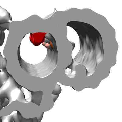
| Title | Subtomogram average (699 axonemal repeats) of isolated Rib72A/B double knock out Tetrahymena cilia showing the lumen of the ciliary doublet microtubule (DMT) | |||||||||
 Map data Map data | Rib72A/BKO Tetrahymena | |||||||||
 Sample Sample |
| |||||||||
| Biological species |   Tetrahymena thermophila (eukaryote) Tetrahymena thermophila (eukaryote) | |||||||||
| Method | subtomogram averaging / cryo EM / Resolution: 40.1 Å | |||||||||
 Authors Authors | Stoddard D / Zhao Y / Bayless B / Gui L / Louka P / Dave D / Surwayanshi S / Tomasi R / Dupuis-Williams P / Baroud C ...Stoddard D / Zhao Y / Bayless B / Gui L / Louka P / Dave D / Surwayanshi S / Tomasi R / Dupuis-Williams P / Baroud C / Gaertig J / Winey M / Nicastro D | |||||||||
 Citation Citation | Journal: Mol Biol Cell / Year: 2018 Title: Tetrahymena RIB72A and RIB72B are microtubule inner proteins in the ciliary doublet microtubules. Authors: Daniel Stoddard / Ying Zhao / Brian A Bayless / Long Gui / Panagiota Louka / Drashti Dave / Swati Suryawanshi / Raphaël F-X Tomasi / Pascale Dupuis-Williams / Charles N Baroud / Jacek ...Authors: Daniel Stoddard / Ying Zhao / Brian A Bayless / Long Gui / Panagiota Louka / Drashti Dave / Swati Suryawanshi / Raphaël F-X Tomasi / Pascale Dupuis-Williams / Charles N Baroud / Jacek Gaertig / Mark Winey / Daniela Nicastro /   Abstract: Doublet and triplet microtubules are essential and highly stable core structures of centrioles, basal bodies, cilia, and flagella. In contrast to dynamic cytoplasmic micro-tubules, their luminal ...Doublet and triplet microtubules are essential and highly stable core structures of centrioles, basal bodies, cilia, and flagella. In contrast to dynamic cytoplasmic micro-tubules, their luminal surface is coated with regularly arranged microtubule inner proteins (MIPs). However, the protein composition and biological function(s) of MIPs remain poorly understood. Using genetic, biochemical, and imaging techniques, we identified Tetrahymena RIB72A and RIB72B proteins as ciliary MIPs. Fluorescence imaging of tagged RIB72A and RIB72B showed that both proteins colocalize to Tetrahymena cilia and basal bodies but assemble independently. Cryoelectron tomography of RIB72A and/or RIB72B knockout strains revealed major structural defects in the ciliary A-tubule involving MIP1, MIP4, and MIP6 structures. The defects of individual mutants were complementary in the double mutant. All mutants had reduced swimming speed and ciliary beat frequencies, and high-speed video imaging revealed abnormal highly curved cilia during power stroke. Our results show that RIB72A and RIB72B are crucial for the structural assembly of ciliary A-tubule MIPs and are important for proper ciliary motility. | |||||||||
| History |
|
- Structure visualization
| Movie |
 Movie viewer Movie viewer |
|---|---|
| Structure viewer | EM map: SurfViewMolmilJmol/JSmol |
| Supplemental images |
- Downloads & links
-EMDB archive
| Map data | emd_7805.map.gz | 3.2 MB | EMDB map data format | |
|---|---|---|---|---|
| Header (meta data) | emd-7805-v30.xmlemd-7805.xml | 12.6 KB 12.6 KB | Display Display | EMDB header |
| Images |  emd_7805.png emd_7805.png | 65.6 KB | ||
| Archive directory |  http://ftp.pdbj.org/pub/emdb/structures/EMD-7805ftp://ftp.pdbj.org/pub/emdb/structures/EMD-7805 http://ftp.pdbj.org/pub/emdb/structures/EMD-7805ftp://ftp.pdbj.org/pub/emdb/structures/EMD-7805 | HTTPS FTP |
-Related structure data


-Links
| EMDB pages | EMDB (EBI/PDBe) / EMDataResource |
|---|
-Map
| File | Download / File: emd_7805.map.gz / Format: CCP4 / Size: 3.8 MB / Type: IMAGE STORED AS FLOATING POINT NUMBER (4 BYTES) | ||||||||||||||||||||||||||||||||||||||||||||||||||||||||||||
|---|---|---|---|---|---|---|---|---|---|---|---|---|---|---|---|---|---|---|---|---|---|---|---|---|---|---|---|---|---|---|---|---|---|---|---|---|---|---|---|---|---|---|---|---|---|---|---|---|---|---|---|---|---|---|---|---|---|---|---|---|---|
| Annotation | Rib72A/BKO Tetrahymena | ||||||||||||||||||||||||||||||||||||||||||||||||||||||||||||
| Projections & slices | Image control
Images are generated by Spider. | ||||||||||||||||||||||||||||||||||||||||||||||||||||||||||||
| Voxel size | X=Y=Z: 10.77 Å | ||||||||||||||||||||||||||||||||||||||||||||||||||||||||||||
| Density |
| ||||||||||||||||||||||||||||||||||||||||||||||||||||||||||||
| Symmetry | Space group: 1 | ||||||||||||||||||||||||||||||||||||||||||||||||||||||||||||
| Details | EMDB XML:
CCP4 map header:
| ||||||||||||||||||||||||||||||||||||||||||||||||||||||||||||
 Z (Sec.)
 Y (Row.)
 X (Col.)


-Supplemental data
- Sample components
-Entire : Subtomogram average (699 axonemal repeats) of isolated Rib72A/B d...
| Entire | Name: Subtomogram average (699 axonemal repeats) of isolated Rib72A/B double knock out Tetrahymena cilia showing the lumen of the ciliary doublet microtubule (DMT) |
|---|---|
| Components |
|
-Supramolecule #1: Subtomogram average (699 axonemal repeats) of isolated Rib72A/B d...
| Supramolecule | Name: Subtomogram average (699 axonemal repeats) of isolated Rib72A/B double knock out Tetrahymena cilia showing the lumen of the ciliary doublet microtubule (DMT) type: organelle_or_cellular_component / ID: 1 / Parent: 0 |
|---|---|
| Source (natural) | Organism: Tetrahymena thermophila (eukaryote) / Strain: Rib72A/B KO |
-Experimental details
-Structure determination
| Method | cryo EM |
|---|---|
 Processing Processing | subtomogram averaging |
| Aggregation state | tissue |
-Sample preparation
| Buffer | pH: 7.4 Component:
Details: Solutions were freshly made before use | ||||||||||||||||||
|---|---|---|---|---|---|---|---|---|---|---|---|---|---|---|---|---|---|---|---|
| Grid | Model: Quantifoil R2/2 / Material: COPPER / Mesh: 200 / Support film - Material: CARBON / Support film - topology: HOLEY / Pretreatment - Type: GLOW DISCHARGE / Pretreatment - Atmosphere: AIR / Pretreatment - Pressure: 101.325 kPa | ||||||||||||||||||
| Vitrification | Cryogen name: ETHANE / Chamber temperature: 100 K / Instrument: HOMEMADE PLUNGER Details: back-side blotting for 1.5-2.5 seconds before plunging. |
- Electron microscopy
| Microscope | FEI TECNAI F30 |
|---|---|
| Specialist optics | Energy filter - Name: GIF 2000 / Energy filter - Lower energy threshold: 0 eV / Energy filter - Upper energy threshold: 20 eV |
| Image recording | Film or detector model: GATAN ULTRASCAN 1000 (2k x 2k) / Average electron dose: 1.5 e/Å2 |
| Electron beam | Acceleration voltage: 300 kV / Electron source:  FIELD EMISSION GUN FIELD EMISSION GUN |
| Electron optics | Calibrated defocus max: 8.0 µm / Calibrated magnification: 13500 / Illumination mode: FLOOD BEAM / Imaging mode: BRIGHT FIELD / Nominal defocus min: 6.0 µm |
| Sample stage | Specimen holder model: GATAN LIQUID NITROGEN / Cooling holder cryogen: NITROGEN |
| Experimental equipment |  Model: Tecnai F30 / Image courtesy: FEI Company |
-Image processing
| Final reconstruction | Applied symmetry - Point group: C1 (asymmetric) / Resolution.type: BY AUTHOR / Resolution: 40.1 Å / Resolution method: FSC 0.5 CUT-OFF / Software - Name: IMOD / Number subtomograms used: 699 |
|---|---|
| Extraction | Number tomograms: 5 / Number images used: 699 / Software - Name: MATLAB |
| Final angle assignment | Type: ANGULAR RECONSTITUTION / Software - Name: PEET |
