[English] 日本語
Yorodumi
- EMDB-77973: pointed end of actin filament with one Lmod2 molecule bound -

+
Open data


ID or keywords:

Loading...

-
Basic information

Entry
Database: EMDB / ID: EMD-77973
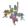
Titlepointed end of actin filament with one Lmod2 molecule bound
Map datapointed end of actin filament with one Lmod2 molecule bound
Sample
  • Complex: Actin, alpha skeletal muscle
    • Complex: Leiomodin-2
Keywordscryo-EM / actin / Lmod2 / actin filament / pointed end / motor protein
Biological speciesOryctolagus cuniculus (rabbit) / Mus musculus (house mouse)
Methodsingle particle reconstruction / cryo EM / Resolution: 8.5 Å
AuthorsRisi CM / Galkin VE
Funding support United States, 2 items
OrganizationGrant numberCountry
National Institutes of Health/National Institute of General Medical Sciences (NIH/NIGMS)R01GM120137 United States
National Institutes of Health/National Heart, Lung, and Blood Institute (NIH/NHLBI)R01 HL160966 United States
CitationJournal: J Mol Cell Cardiol / Year: 2026
Title: Structure of the actively growing actin filament pointed end facilitated by Leiomodin-2.
Authors: Cristina M Risi / Tania M Larrinaga / Alla S Kostyukova / Carol C Gregorio / Vitold E Galkin /
Abstract: Cardiac contraction depends on synchronized interactions between myosin-based thick filaments and actin-based thin filaments (TFs). Precise regulation of TF length is vital for cardiac function, as ...Cardiac contraction depends on synchronized interactions between myosin-based thick filaments and actin-based thin filaments (TFs). Precise regulation of TF length is vital for cardiac function, as any alteration in length leads to severe myopathies. Actin filaments form the backbone of the TF and have two unequal ends - fast-growing barbed and slow-growing pointed. In muscle, the barbed end is capped at the Z-line, so the thin filament can elongate only from the pointed end. Leiomodin-2 (Lmod2) promotes actin nucleation and possesses a unique C-terminal extension (CTE) that is important for actin nucleation and binds to the sides of mature TFs. Single-molecule studies have shown that Lmod2 remains bound processively to the growing pointed end. We employed cryo-electron microscopy to visualize the structure of actively growing pointed ends in the presence of Lmod2 from G-actin complexed with profilin (profilactin). We show that Lmod2's leucine-rich repeat domain (LRR) stabilizes terminal actin subunits by binding across the helical groove of actin on one or both actin strands. LRR binding pushes the terminal actins outward from their ideal positions in the actin filament, introducing strain at the pointed end that squeezes Lmod2 from the filament's exterior, thereby maintaining the concentration of Lmod2 required for further elongation. Further, we suggest that the unique Lmod2 CTE may stabilize Lmod2 binding to the actively growing pointed end.
History
DepositionJul 7, 2026-
Header (metadata) releaseOct 21, 2026-
Map releaseOct 21, 2026-
UpdateOct 21, 2026-
Current statusOct 21, 2026Processing site: RCSB / Status: Released

-
Structure visualization

Supplemental images

Downloads & links

-
Map

FileDownload / File: emd_77973.map.gz / Format: CCP4 / Size: 244.1 MB / Type: IMAGE STORED AS FLOATING POINT NUMBER (4 BYTES)
Annotationpointed end of actin filament with one Lmod2 molecule bound
Projections & slices

Image control

Size
Brightness
Contrast
Others
AxesZ (Sec.)Y (Row.)X (Col.)
1.36 Å/pix.
x 400 pix.
= 542.4 Å
1.36 Å/pix.
x 400 pix.
= 542.4 Å
1.36 Å/pix.
x 400 pix.
= 542.4 Å

Surface

Projections

Slices (1/3)

Slices (1/2)

Slices (2/3)

Images are generated by Spider.

Voxel sizeX=Y=Z: 1.356 Å
Density
Contour LevelBy AUTHOR: 0.7
Minimum - Maximum-3.1549873 - 5.739662
Average (Standard dev.)0.00027118673 (±0.14717339)
SymmetrySpace group: 1
Details

EMDB XML:

Map geometry
Axis orderXYZ
Origin000
Dimensions400400400
Spacing400400400
CellA=B=C: 542.39996 Å
α=β=γ: 90.0 °

-
Supplemental data

-
Half map: Half Map A

Fileemd_77973_half_map_1.map
AnnotationHalf Map A
Projections & Slices
AxesZYX

Projections

Slices (1/2)
Density Histograms

-
Half map: Half Map B

Fileemd_77973_half_map_2.map
AnnotationHalf Map B
Projections & Slices
AxesZYX

Projections

Slices (1/2)
Density Histograms

-
Sample components

-
Entire : Actin, alpha skeletal muscle

EntireName: Actin, alpha skeletal muscle
Components
  • Complex: Actin, alpha skeletal muscle
    • Complex: Leiomodin-2

-
Supramolecule #1: Actin, alpha skeletal muscle

SupramoleculeName: Actin, alpha skeletal muscle / type: complex / ID: 1 / Parent: 0
Source (natural)Organism: Oryctolagus cuniculus (rabbit)

-
Supramolecule #2: Leiomodin-2

SupramoleculeName: Leiomodin-2 / type: complex / ID: 2 / Parent: 1
Source (natural)Organism: Mus musculus (house mouse)

-
Experimental details

-
Structure determination

Methodcryo EM
Processingsingle particle reconstruction
Aggregation statefilament

-
Sample preparation

BufferpH: 7.5
GridModel: EMS Lacey Carbon / Material: COPPER / Mesh: 300 / Support film - Material: CARBON / Support film - topology: LACEY / Pretreatment - Type: GLOW DISCHARGE / Pretreatment - Time: 45 sec.
VitrificationCryogen name: ETHANE

-
Electron microscopy

MicroscopeTFS KRIOS
Image recordingFilm or detector model: GATAN K3 (6k x 4k) / Number grids imaged: 4 / Number real images: 11689 / Average electron dose: 34.0 e/Å2 / Details: 40 frames taken per image
Electron beamAcceleration voltage: 300 kV / Electron source: FIELD EMISSION GUN
Electron opticsIllumination mode: FLOOD BEAM / Imaging mode: BRIGHT FIELD / Nominal defocus max: 3.5 µm / Nominal defocus min: 0.5 µm
Sample stageSpecimen holder model: FEI TITAN KRIOS AUTOGRID HOLDER / Cooling holder cryogen: NITROGEN
Experimental equipment
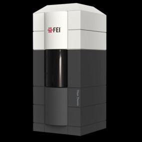
Model: Titan Krios / Image courtesy: FEI Company

+
Image processing

Particle selectionNumber selected: 21712
CTF correctionSoftware - Name: CTFFIND (ver. 4) / Type: PHASE FLIPPING AND AMPLITUDE CORRECTION
Startup modelType of model: PDB ENTRY
PDB model - PDB ID:
Final reconstructionAlgorithm: BACK PROJECTION / Resolution.type: BY AUTHOR / Resolution: 8.5 Å / Resolution method: FSC 0.143 CUT-OFF / Software - Name: RELION (ver. 4.0) / Details: Map low-pass filtered to 9.5 Angstroms / Number images used: 1992
Initial angle assignmentType: MAXIMUM LIKELIHOOD / Software - Name: RELION (ver. 4)
Final angle assignmentType: MAXIMUM LIKELIHOOD / Software - Name: RELION (ver. 4)
Final 3D classificationNumber classes: 3 / Software - Name: RELION (ver. 4)
FSC plot (resolution estimation)

-
Atomic model buiding 1

Initial model
PDB IDChain

source_name: PDB, initial_model_type: experimental model

source_name: PDB, initial_model_type: experimental model
RefinementSpace: REAL / Protocol: FLEXIBLE FIT

+
About Yorodumi

-
News

-
Feb 9, 2022. New format data for meta-information of EMDB entries

New format data for meta-information of EMDB entries

  • Version 3 of the EMDB header file is now the official format.
  • The previous official version 1.9 will be removed from the archive.

Related info.:EMDB header

External links:wwPDB to switch to version 3 of the EMDB data model

-
Aug 12, 2020. Covid-19 info

Covid-19 info

URL: https://pdbj.org/emnavi/covid19.php

New page: Covid-19 featured information page in EM Navigator.

Related info.:Covid-19 info / Mar 5, 2020. Novel coronavirus structure data

+
Mar 5, 2020. Novel coronavirus structure data

Novel coronavirus structure data

Related info.:Yorodumi Speices / Aug 12, 2020. Covid-19 info

External links:COVID-19 featured content - PDBj / Molecule of the Month (242):Coronavirus Proteases

+
Jan 31, 2019. EMDB accession codes are about to change! (news from PDBe EMDB page)

EMDB accession codes are about to change! (news from PDBe EMDB page)

  • The allocation of 4 digits for EMDB accession codes will soon come to an end. Whilst these codes will remain in use, new EMDB accession codes will include an additional digit and will expand incrementally as the available range of codes is exhausted. The current 4-digit format prefixed with “EMD-” (i.e. EMD-XXXX) will advance to a 5-digit format (i.e. EMD-XXXXX), and so on. It is currently estimated that the 4-digit codes will be depleted around Spring 2019, at which point the 5-digit format will come into force.
  • The EM Navigator/Yorodumi systems omit the EMD- prefix.

Related info.:Q: What is EMD? / ID/Accession-code notation in Yorodumi/EM Navigator

External links:EMDB Accession Codes are Changing Soon! / Contact to PDBj

+
Jul 12, 2017. Major update of PDB

Major update of PDB

  • wwPDB released updated PDB data conforming to the new PDBx/mmCIF dictionary.
  • This is a major update changing the version number from 4 to 5, and with Remediation, in which all the entries are updated.
  • In this update, many items about electron microscopy experimental information are reorganized (e.g. em_software).
  • Now, EM Navigator and Yorodumi are based on the updated data.

External links:wwPDB Remediation / Enriched Model Files Conforming to OneDep Data Standards Now Available in the PDB FTP Archive

-
Yorodumi

Thousand views of thousand structures

  • Yorodumi is a browser for structure data from EMDB, PDB, SASBDB, etc.
  • This page is also the successor to EM Navigator detail page, and also detail information page/front-end page for Omokage search.
  • The word "yorodu" (or yorozu) is an old Japanese word meaning "ten thousand". "mi" (miru) is to see.

Related info.:EMDB / PDB / SASBDB / Comparison of 3 databanks / Yorodumi Search / Aug 31, 2016. New EM Navigator & Yorodumi / Yorodumi Papers / Jmol/JSmol / Function and homology information / Changes in new EM Navigator and Yorodumi

Read more