 Movie
Movie Controller

[English] 日本語
 Yorodumi
- EMDB-7778: Whole-population, doublet-microtubule map from a Drosophila melan... -
+ Open data

- Basic information
| Entry | Database: EMDB / ID: EMD-7778 | |||||||||
|---|---|---|---|---|---|---|---|---|---|---|
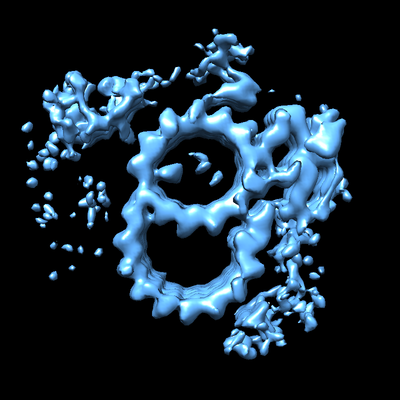
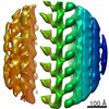
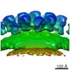
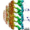
| Title | Whole-population, doublet-microtubule map from a Drosophila melanogaster S2 cell centriole | |||||||||
 Map data Map data | primary map | |||||||||
 Sample Sample |
| |||||||||
| Biological species |  | |||||||||
| Method | subtomogram averaging / cryo EM / Resolution: 30.0 Å | |||||||||
 Authors Authors | Agard DA / Greenan GA | |||||||||
 Citation Citation | Journal: Elife / Year: 2018 Title: Insights into centriole geometry revealed by cryotomography of doublet and triplet centrioles. Authors: Garrett A Greenan / Bettina Keszthelyi / Ronald D Vale / David A Agard /  Abstract: Centrioles are cylindrical assemblies comprised of 9 singlet, doublet, or triplet microtubules, essential for the formation of motile and sensory cilia. While the structure of the cilium is being ...Centrioles are cylindrical assemblies comprised of 9 singlet, doublet, or triplet microtubules, essential for the formation of motile and sensory cilia. While the structure of the cilium is being defined at increasing resolution, centriolar structure remains poorly understood. Here, we used electron cryo-tomography to determine the structure of mammalian (triplet) and (doublet) centrioles. Mammalian centrioles have two distinct domains: a 200 nm proximal core region connected by A-C linkers, and a distal domain where the C-tubule is incomplete and a pair of novel linkages stabilize the assembly producing a geometry more closely resembling the ciliary axoneme. centrioles resemble the mammalian core, but with their doublet microtubules linked through the A tubules. The commonality of core-region length, and the abrupt transition in mammalian centrioles, suggests a conserved length-setting mechanism. The unexpected linker diversity suggests how unique centriolar architectures arise in different tissues and organisms. | |||||||||
| History |
|
- Structure visualization
| Movie |
 Movie viewer Movie viewer |
|---|---|
| Structure viewer | EM map: SurfViewMolmilJmol/JSmol |
| Supplemental images |
- Downloads & links
-EMDB archive
| Map data | emd_7778.map.gz | 1.1 MB | EMDB map data format | |
|---|---|---|---|---|
| Header (meta data) | emd-7778-v30.xmlemd-7778.xml | 7.1 KB 7.1 KB | Display Display | EMDB header |
| Images |  emd_7778.png emd_7778.png | 117.6 KB | ||
| Archive directory |  http://ftp.pdbj.org/pub/emdb/structures/EMD-7778ftp://ftp.pdbj.org/pub/emdb/structures/EMD-7778 http://ftp.pdbj.org/pub/emdb/structures/EMD-7778ftp://ftp.pdbj.org/pub/emdb/structures/EMD-7778 | HTTPS FTP |
-Validation report
| Summary document | emd_7778_validation.pdf.gz | 77.2 KB | Display | EMDB validaton report |
|---|---|---|---|---|
| Full document | emd_7778_full_validation.pdf.gz | 76.3 KB | Display | |
| Data in XML | emd_7778_validation.xml.gz | 493 B | Display | |
| Arichive directory | https://ftp.pdbj.org/pub/emdb/validation_reports/EMD-7778ftp://ftp.pdbj.org/pub/emdb/validation_reports/EMD-7778 | HTTPS FTP |
-Related structure data


-Links
| EMDB pages | EMDB (EBI/PDBe) / EMDataResource |
|---|
-Map
| File | Download / File: emd_7778.map.gz / Format: CCP4 / Size: 12.9 MB / Type: IMAGE STORED AS FLOATING POINT NUMBER (4 BYTES) | ||||||||||||||||||||||||||||||||||||||||||||||||||||||||||||
|---|---|---|---|---|---|---|---|---|---|---|---|---|---|---|---|---|---|---|---|---|---|---|---|---|---|---|---|---|---|---|---|---|---|---|---|---|---|---|---|---|---|---|---|---|---|---|---|---|---|---|---|---|---|---|---|---|---|---|---|---|---|
| Annotation | primary map | ||||||||||||||||||||||||||||||||||||||||||||||||||||||||||||
| Projections & slices | Image control
Images are generated by Spider. | ||||||||||||||||||||||||||||||||||||||||||||||||||||||||||||
| Voxel size | X=Y=Z: 8.18 Å | ||||||||||||||||||||||||||||||||||||||||||||||||||||||||||||
| Density |
| ||||||||||||||||||||||||||||||||||||||||||||||||||||||||||||
| Symmetry | Space group: 1 | ||||||||||||||||||||||||||||||||||||||||||||||||||||||||||||
| Details | EMDB XML:
CCP4 map header:
| ||||||||||||||||||||||||||||||||||||||||||||||||||||||||||||
 Z (Sec.)
 Y (Row.)
 X (Col.)


-Supplemental data
- Sample components
-Entire : Whole-population, doublet-microtubule map from a Drosophila melan...
| Entire | Name: Whole-population, doublet-microtubule map from a Drosophila melanogaster S2 cell centriole |
|---|---|
| Components |
|
-Supramolecule #1: Whole-population, doublet-microtubule map from a Drosophila melan...
| Supramolecule | Name: Whole-population, doublet-microtubule map from a Drosophila melanogaster S2 cell centriole type: complex / ID: 1 / Parent: 0 |
|---|---|
| Source (natural) | Organism: |
-Experimental details
-Structure determination
| Method | cryo EM |
|---|---|
 Processing Processing | subtomogram averaging |
| Aggregation state | cell |
-Sample preparation
| Buffer | pH: 7.2 |
|---|---|
| Vitrification | Cryogen name: ETHANE |
- Electron microscopy
| Microscope | FEI POLARA 300 |
|---|---|
| Image recording | Film or detector model: GATAN K2 SUMMIT (4k x 4k) / Average electron dose: 8.0 e/Å2 |
| Electron beam | Acceleration voltage: 300 kV / Electron source:  FIELD EMISSION GUN FIELD EMISSION GUN |
| Electron optics | Illumination mode: OTHER / Imaging mode: BRIGHT FIELD |
| Experimental equipment |  Model: Tecnai Polara / Image courtesy: FEI Company |
-Image processing
| Final reconstruction | Applied symmetry - Point group: C1 (asymmetric) / Resolution.type: BY AUTHOR / Resolution: 30.0 Å / Resolution method: FSC 0.143 CUT-OFF / Number subtomograms used: 30 |
|---|---|
| Extraction | Number tomograms: 30 / Number images used: 2300 |
| Final angle assignment | Type: ANGULAR RECONSTITUTION |
