Movie
Movie Controller


+ Open data

- Basic information
| Entry | Database: EMDB / ID: EMD-7471 | |||||||||
|---|---|---|---|---|---|---|---|---|---|---|
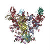
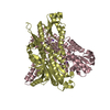
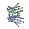
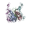
| Title | QA013.2 Fab fragment bound to BG505 T332N SOSIP.664 trimer | |||||||||
 Map data Map data | QA013.2-Fab fragment bound to BG505 T332N SOSIP.664 trimer | |||||||||
 Sample Sample |
| |||||||||
| Biological species |   Human immunodeficiency virus / Human immunodeficiency virus /  Homo sapiens (human) Homo sapiens (human) | |||||||||
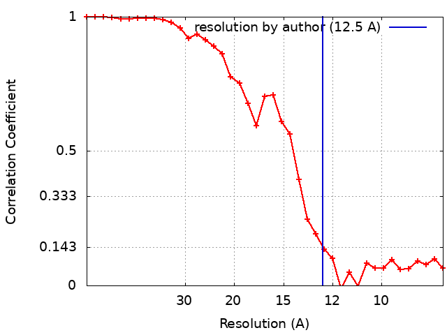
| Method | single particle reconstruction / negative staining / Resolution: 12.5 Å | |||||||||
 Authors Authors | Williams JA / Lee KK | |||||||||
 Citation Citation | Journal: Cell Rep / Year: 2018 Title: Superinfection Drives HIV Neutralizing Antibody Responses from Several B Cell Lineages that Contribute to a Polyclonal Repertoire. Authors: Katherine L Williams / Bingjie Wang / Dana Arenz / James A Williams / Adam S Dingens / Valerie Cortez / Cassandra A Simonich / Stephanie Rainwater / Dara A Lehman / Kelly K Lee / Julie Overbaugh /  Abstract: Eliciting broad and potent HIV-specific neutralizing antibody responses represents the holy grail of HIV vaccine efforts. Data from singly infected individuals with broad and potent plasma ...Eliciting broad and potent HIV-specific neutralizing antibody responses represents the holy grail of HIV vaccine efforts. Data from singly infected individuals with broad and potent plasma neutralizing activity targeting one epitope have guided our understanding of how these responses develop. However, far less is known about responses developed by superinfected individuals who acquire two distinct HIV strains. Here, we isolated HIV-specific mAbs from a superinfected individual with a broad plasma response. In this superinfection case, neutralizing activity resulted from multiple distinct B cell lineages that arose in response to either the initial or the superinfecting virus, including an antibody that targets the N332 supersite. This nAb, QA013.2, was specific to the superinfecting virus and was associated with eventual reemergence of the initial infecting virus. The complex dynamic between viruses in superinfection may drive development of a unique collection of polyclonal nAbs that present a higher barrier to escape than monoclonal responses. | |||||||||
| History |
|
- Structure visualization
| Movie |
 Movie viewer Movie viewer |
|---|---|
| Structure viewer | EM map: SurfViewMolmilJmol/JSmol |
| Supplemental images |
 UCSF Chimera
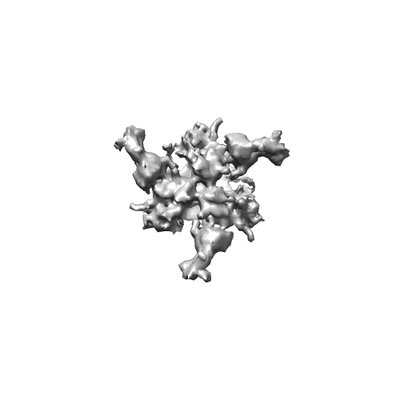
- Downloads & links
-EMDB archive
| Map data | emd_7471.map.gz | 552.8 KB | EMDB map data format | |
|---|---|---|---|---|
| Header (meta data) | emd-7471-v30.xmlemd-7471.xml | 11 KB 11 KB | Display Display | EMDB header |
| FSC (resolution estimation) | emd_7471_fsc.xml | 3.6 KB | Display | FSC data file |
| Images |  emd_7471.png emd_7471.png | 16.6 KB | ||
| Archive directory |  http://ftp.pdbj.org/pub/emdb/structures/EMD-7471ftp://ftp.pdbj.org/pub/emdb/structures/EMD-7471 http://ftp.pdbj.org/pub/emdb/structures/EMD-7471ftp://ftp.pdbj.org/pub/emdb/structures/EMD-7471 | HTTPS FTP |
-Related structure data
| Similar structure data |
|---|


-Links
| EMDB pages | EMDB (EBI/PDBe) / EMDataResource |
|---|
-Map
| File | Download / File: emd_7471.map.gz / Format: CCP4 / Size: 2.1 MB / Type: IMAGE STORED AS FLOATING POINT NUMBER (4 BYTES) | ||||||||||||||||||||||||||||||||||||||||||||||||||||||||||||||||||||
|---|---|---|---|---|---|---|---|---|---|---|---|---|---|---|---|---|---|---|---|---|---|---|---|---|---|---|---|---|---|---|---|---|---|---|---|---|---|---|---|---|---|---|---|---|---|---|---|---|---|---|---|---|---|---|---|---|---|---|---|---|---|---|---|---|---|---|---|---|---|
| Annotation | QA013.2-Fab fragment bound to BG505 T332N SOSIP.664 trimer | ||||||||||||||||||||||||||||||||||||||||||||||||||||||||||||||||||||
| Projections & slices | Image control
Images are generated by Spider. | ||||||||||||||||||||||||||||||||||||||||||||||||||||||||||||||||||||
| Voxel size | X=Y=Z: 2.07 Å | ||||||||||||||||||||||||||||||||||||||||||||||||||||||||||||||||||||
| Density |
| ||||||||||||||||||||||||||||||||||||||||||||||||||||||||||||||||||||
| Symmetry | Space group: 1 | ||||||||||||||||||||||||||||||||||||||||||||||||||||||||||||||||||||
| Details | EMDB XML:
CCP4 map header:
| ||||||||||||||||||||||||||||||||||||||||||||||||||||||||||||||||||||
 Z (Sec.)
 Y (Row.)
 X (Col.)


-Supplemental data
- Sample components
-Entire : Complex of QA013.2 Fab bound to BG505 T332N SOSIP.664 trimer
| Entire | Name: Complex of QA013.2 Fab bound to BG505 T332N SOSIP.664 trimer |
|---|---|
| Components |
|
-Supramolecule #1: Complex of QA013.2 Fab bound to BG505 T332N SOSIP.664 trimer
| Supramolecule | Name: Complex of QA013.2 Fab bound to BG505 T332N SOSIP.664 trimer type: complex / ID: 1 / Parent: 0 Details: Fab fragment generated by papain digestion of IgG antibody. SOSIP.664 Env trimer recombinantly expressed in HEK-293F cells. |
|---|---|
| Source (natural) | Organism: Human immunodeficiency virus |
| Molecular weight | Theoretical: 360 KDa |
-Supramolecule #2: QA0132 Fab fragment
| Supramolecule | Name: QA0132 Fab fragment / type: complex / ID: 2 / Parent: 1 / Details: Fab fragment generated by papain digestion of IgG |
|---|---|
| Source (natural) | Organism: Homo sapiens (human) |
| Recombinant expression | Organism: Homo sapiens (human) / Recombinant cell: Human embryonic kidney / Recombinant plasmid: pPPI4 |
-Supramolecule #3: BG505 T332N SOSIP.664 trimer
| Supramolecule | Name: BG505 T332N SOSIP.664 trimer / type: complex / ID: 3 / Parent: 1 |
|---|
-Experimental details
-Structure determination
| Method | negative staining |
|---|---|
 Processing Processing | single particle reconstruction |
| Aggregation state | particle |
-Sample preparation
| Concentration | .020 mg/mL |
|---|---|
| Buffer | pH: 7.5 |
| Staining | Type: NEGATIVE / Material: Methylamine Tungstate |
| Grid | Model: C-flat / Material: COPPER / Mesh: 300 / Support film - Material: CARBON / Support film - topology: CONTINUOUS / Pretreatment - Type: GLOW DISCHARGE |
- Electron microscopy
| Microscope | FEI TECNAI 12 |
|---|---|
| Image recording | Film or detector model: GATAN ULTRASCAN 4000 (4k x 4k) / Number grids imaged: 1 / Number real images: 146 / Average exposure time: 1.0 sec. / Average electron dose: 27.0 e/Å2 |
| Electron beam | Acceleration voltage: 120 kV / Electron source: LAB6 |
| Electron optics | Illumination mode: FLOOD BEAM / Imaging mode: BRIGHT FIELD / Nominal defocus max: 3.0 µm / Nominal defocus min: 2.0 µm / Nominal magnification: 52000 |
| Sample stage | Specimen holder model: SIDE ENTRY, EUCENTRIC |