 Movie
Movie Controller

[English] 日本語
 Yorodumi
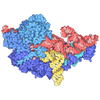
- EMDB-73602: Babesia divergens ribosome structure by single-particle cryo-EM (... -
+ Open data

- Basic information
| Entry |  | |||||||||
|---|---|---|---|---|---|---|---|---|---|---|
| Title | Babesia divergens ribosome structure by single-particle cryo-EM (3D class1, A-, P-, and E-site tRNAs and mRNA) | |||||||||
 Map data Map data | ||||||||||
 Sample Sample |
| |||||||||
 Keywords Keywords | tRNAs / RNA modification / 80S / RIBOSOME | |||||||||
| Function / homology |  Function and homology information Function and homology informationpreribosome / 90S preribosome / protein-RNA complex assembly / translation regulator activity / ribosomal large subunit biogenesis / maturation of SSU-rRNA from tricistronic rRNA transcript (SSU-rRNA, 5.8S rRNA, LSU-rRNA) / maturation of LSU-rRNA from tricistronic rRNA transcript (SSU-rRNA, 5.8S rRNA, LSU-rRNA) / maturation of SSU-rRNA / small-subunit processome / chloroplast ...preribosome / 90S preribosome / protein-RNA complex assembly / translation regulator activity / ribosomal large subunit biogenesis / maturation of SSU-rRNA from tricistronic rRNA transcript (SSU-rRNA, 5.8S rRNA, LSU-rRNA) / maturation of LSU-rRNA from tricistronic rRNA transcript (SSU-rRNA, 5.8S rRNA, LSU-rRNA) / maturation of SSU-rRNA / small-subunit processome / chloroplast / kinase activity / rRNA processing / large ribosomal subunit / ribosomal small subunit assembly / ribosome binding / ribosomal small subunit biogenesis / ribosome biogenesis / 5S rRNA binding / ribosomal large subunit assembly / small ribosomal subunit / cytosolic small ribosomal subunit / small ribosomal subunit rRNA binding / large ribosomal subunit rRNA binding / cytosolic large ribosomal subunit / cytoplasmic translation / negative regulation of translation / rRNA binding / ribosome / translation / structural constituent of ribosome / ribonucleoprotein complex / mRNA binding / nucleolus / RNA binding / zinc ion binding / nucleus / cytosol / cytoplasm Similarity search - Function | |||||||||
| Biological species |  Babesia divergens (eukaryote) Babesia divergens (eukaryote) | |||||||||
| Method | single particle reconstruction / cryo EM / Resolution: 3.1 Å | |||||||||
 Authors Authors | Gutierrez-Vargas C / Izhaki-Tavor LS / Leger-Abraham M | |||||||||
| Funding support |  United States, 2 items United States, 2 items
| |||||||||
 Citation Citation | Journal: bioRxiv / Year: 2025 Title: Ribosomal Architecture and rRNA Modification Landscape in the Tick-Borne Parasite . Authors: Cristina Gutierrez-Vargas / Lee S Izhaki-Tavor / Diana Calvopina-Chavez / Caroline D Keroack / Pablo Copello / Manoj T Duraisingh / Mélissa Léger-Abraham / Abstract: is a tick-borne intracellular apicomplexan parasite responsible for diseases ranging from mild to fatal, with a broadening geographic distribution. Due to the complex life cycle of species, their ... is a tick-borne intracellular apicomplexan parasite responsible for diseases ranging from mild to fatal, with a broadening geographic distribution. Due to the complex life cycle of species, their survival depends on the precise control of gene expression, which is primarily regulated by epigenetic, transcriptional, and post-transcriptional mechanisms. High-resolution structural information on key components of the translation machinery, such as ribosomes, could aid in the development of antiparasitic drugs. Here, we report cryo-EM ribosome structures (2.6 Å) from the tick-borne apicomplexan pathogen , showing associated tRNAs, an mRNA fragment, and RACK1, a signaling scaffold crucial to translation regulation. Density map analysis displays ribosome regions at atomic resolution (1.7 Å), which, when combined with nanopore sequencing, enabled the comprehensive identification of rRNA modifications, including modifications unreported in other organisms. The new rRNA modifications localize not only to the reduced rRNA expansion segments but also to functionally essential ribosomal sites, uncovering new avenues for therapeutic intervention against babesiosis. | |||||||||
| History |
|
- Structure visualization
| Supplemental images |
|---|

- Downloads & links
-EMDB archive
| Map data | emd_73602.map.gz | 307.4 MB | EMDB map data format | |
|---|---|---|---|---|
| Header (meta data) | emd-73602-v30.xmlemd-73602.xml | 124.4 KB 124.4 KB | Display Display | EMDB header |
| Images |  emd_73602.png emd_73602.png | 125.4 KB | ||
| Filedesc metadata | emd-73602.cif.gz | 21 KB | ||
| Others | emd_73602_additional_1.map.gz | 36.9 MB | ||
| Archive directory |  http://ftp.pdbj.org/pub/emdb/structures/EMD-73602ftp://ftp.pdbj.org/pub/emdb/structures/EMD-73602 http://ftp.pdbj.org/pub/emdb/structures/EMD-73602ftp://ftp.pdbj.org/pub/emdb/structures/EMD-73602 | HTTPS FTP |
-Related structure data
| Related structure data |  9yxbMC  9ygmC C: citing same article ( M: atomic model generated by this map |
|---|---|
| Similar structure data |


-Links
| EMDB pages | EMDB (EBI/PDBe) / EMDataResource |
|---|---|
| Related items in Molecule of the Month |

-Map
| File | Download / File: emd_73602.map.gz / Format: CCP4 / Size: 343 MB / Type: IMAGE STORED AS FLOATING POINT NUMBER (4 BYTES) | ||||||||||||||||||||||||||||||||||||
|---|---|---|---|---|---|---|---|---|---|---|---|---|---|---|---|---|---|---|---|---|---|---|---|---|---|---|---|---|---|---|---|---|---|---|---|---|---|
| Projections & slices | Image control
Images are generated by Spider. | ||||||||||||||||||||||||||||||||||||
| Voxel size | X=Y=Z: 0.825 Å | ||||||||||||||||||||||||||||||||||||
| Density |
| ||||||||||||||||||||||||||||||||||||
| Symmetry | Space group: 1 | ||||||||||||||||||||||||||||||||||||
| Details | EMDB XML:
|
 Z (Sec.)
 Y (Row.)
 X (Col.)


-Supplemental data
-Additional map: #1
| File | emd_73602_additional_1.map | ||||||||||||
|---|---|---|---|---|---|---|---|---|---|---|---|---|---|
| Projections & Slices |
| ||||||||||||
| Density Histograms |


- Sample components
+Entire : 80S ribosome
+Supramolecule #1: 80S ribosome
+Supramolecule #2: 40S
+Supramolecule #3: 60S
+Macromolecule #1: 40S ribosomal protein uS12
+Macromolecule #2: 40S ribosomal protein eS19
+Macromolecule #3: 40S ribosomal protein uS17
+Macromolecule #4: 40S ribosomal protein uS9
+Macromolecule #5: 40S ribosomal protein uS13
+Macromolecule #6: 40S ribosomal protein uS7
+Macromolecule #7: 40S ribosomal protein uS15
+Macromolecule #8: 40S ribosomal protein eS1
+Macromolecule #9: 40S ribosomal protein eS10
+Macromolecule #10: 40S ribosomal protein uS8
+Macromolecule #11: 40S ribosomal protein eS7
+Macromolecule #12: 40S ribosomal protein eS8
+Macromolecule #13: 40S ribosomal protein eS6
+Macromolecule #14: 40S ribosomal protein uS10
+Macromolecule #15: 40S ribosomal protein eS30
+Macromolecule #16: 40S ribosomal protein uS14
+Macromolecule #17: 40S ribosomal protein eS28
+Macromolecule #18: 40S ribosomal protein uS3
+Macromolecule #19: 40S ribosomal protein uS19
+Macromolecule #20: 40S ribosomal protein uS2
+Macromolecule #21: 40S ribosomal protein uS5
+Macromolecule #22: 40S ribosomal protein eS26
+Macromolecule #23: 40S ribosomal protein eS17
+Macromolecule #24: 40S ribosomal protein uS11
+Macromolecule #25: 40S ribosomal protein eS24
+Macromolecule #26: 40S ribosomal protein eS25
+Macromolecule #27: 40S ribosomal protein eS12
+Macromolecule #28: 40S ribosomal protein uS4
+Macromolecule #29: 40S ribosomal protein eS31
+Macromolecule #30: 40S ribosomal protein eS27
+Macromolecule #32: 60S ribosomal protein uL15
+Macromolecule #33: 60S ribosomal protein eL20
+Macromolecule #34: 60S ribosomal protein eL31
+Macromolecule #35: 60S ribosomal protein eL32
+Macromolecule #36: 60S ribosomal protein eL37
+Macromolecule #37: 60S ribosomal protein eL18
+Macromolecule #38: 60S ribosomal protein uL2
+Macromolecule #39: 60S ribosomal protein uL13
+Macromolecule #40: 60S ribosomal protein eL13
+Macromolecule #41: 60S ribosomal protein uL24
+Macromolecule #42: 60S ribosomal protein uL29
+Macromolecule #43: 60S ribosomal protein uL4
+Macromolecule #44: 60S ribosomal protein uL18
+Macromolecule #45: 60S ribosomal protein eL27
+Macromolecule #46: 60S ribosomal protein uL22
+Macromolecule #47: 60S ribosomal protein eL8
+Macromolecule #48: 60S ribosomal protein uL3
+Macromolecule #49: 60S ribosomal protein eL14
+Macromolecule #50: 60S ribosomal protein uL14
+Macromolecule #51: 60S ribosomal protein uL23
+Macromolecule #52: 60S ribosomal protein eL36
+Macromolecule #53: 60S ribosomal protein eL40
+Macromolecule #54: 60S ribosomal protein eL21
+Macromolecule #55: 60S ribosomal protein eL42
+Macromolecule #56: 60S ribosomal protein eL39
+Macromolecule #57: 60S ribosomal protein eL19
+Macromolecule #58: 60S ribosomal protein uL5
+Macromolecule #59: 60S ribosomal protein uL16
+Macromolecule #60: 60S ribosomal protein eL15
+Macromolecule #61: 60S ribosomal protein eL33
+Macromolecule #62: 60S ribosomal protein uL30
+Macromolecule #63: 60S ribosomal protein eL22
+Macromolecule #64: 60S ribosomal protein eL30
+Macromolecule #65: 60S ribosomal protein eL43
+Macromolecule #66: 60S ribosomal protein eL29
+Macromolecule #67: 60S ribosomal protein eL34
+Macromolecule #68: 60S ribosomal protein eL24
+Macromolecule #69: 60S ribosomal protein eL6
+Macromolecule #70: 60S ribosomal protein eL41
+Macromolecule #78: 40S ribosomal protein eS21
+Macromolecule #79: 40S ribosomal protein eS4
+Macromolecule #80: Receptor for activated C kinase 1, RACK1 protein
+Macromolecule #81: 60S ribosomal protein uL6
+Macromolecule #82: 60S ribosomal protein eL38
+Macromolecule #31: 18S ribosomal RNA
+Macromolecule #71: 5S ribosomal RNA
+Macromolecule #72: 5.8S ribosomal RNA
+Macromolecule #73: 28S ribosomal RNA
+Macromolecule #74: E-site tRNA
+Macromolecule #75: P-site tRNA
+Macromolecule #76: A-site tRNA
+Macromolecule #77: mRNA fragment
+Macromolecule #83: MAGNESIUM ION
+Macromolecule #84: POTASSIUM ION
-Experimental details
-Structure determination
| Method | cryo EM |
|---|---|
 Processing Processing | single particle reconstruction |
| Aggregation state | particle |
-Sample preparation
| Buffer | pH: 7.4 Component:
| ||||||||||||||||||
|---|---|---|---|---|---|---|---|---|---|---|---|---|---|---|---|---|---|---|---|
| Grid | Model: Quantifoil R1.2/1.3 / Material: COPPER / Mesh: 300 / Support film - Material: CARBON / Support film - topology: HOLEY / Support film - Film thickness: 12 / Pretreatment - Type: GLOW DISCHARGE / Pretreatment - Time: 30 sec. / Pretreatment - Atmosphere: AIR / Pretreatment - Pressure: 0.037 kPa Details: The grid had an additional ultrathin continous carbon layer (2 nm) | ||||||||||||||||||
| Vitrification | Cryogen name: ETHANE / Chamber humidity: 100 % / Chamber temperature: 277.15 K / Instrument: FEI VITROBOT MARK IV |
- Electron microscopy
| Microscope | TFS KRIOS |
|---|---|
| Software | Name: EPU |
| Image recording | Film or detector model: GATAN K3 (6k x 4k) / Number real images: 12144 / Average exposure time: 1.4 sec. / Average electron dose: 42.0 e/Å2 |
| Electron beam | Acceleration voltage: 300 kV / Electron source:  FIELD EMISSION GUN FIELD EMISSION GUN |
| Electron optics | C2 aperture diameter: 50.0 µm / Illumination mode: FLOOD BEAM / Imaging mode: BRIGHT FIELD / Cs: 2.7 mm / Nominal defocus max: 2.2 µm / Nominal defocus min: 0.8 µm / Nominal magnification: 105000 |
| Sample stage | Specimen holder model: FEI TITAN KRIOS AUTOGRID HOLDER / Cooling holder cryogen: NITROGEN |
| Experimental equipment |  Model: Titan Krios / Image courtesy: FEI Company |
+Image processing
-Atomic model buiding 1
| Initial model |
| ||||||||||||||||||
|---|---|---|---|---|---|---|---|---|---|---|---|---|---|---|---|---|---|---|---|
| Details | Protein chains were built de novo with ModelAngelo. The rRNA chains from PDB 9YGM, which corresponds to the highest-resolution conformational class (3DC3, 80S with E-site tRNA) from the same dataset, were rigid-body fit and remodeled. The complete model then underwent iterative rounds of manual adjustment in Coot and real-space refinement in Phenix. | ||||||||||||||||||
| Refinement | Space: REAL / Protocol: RIGID BODY FIT | ||||||||||||||||||
| Output model | PDB-9yxb: |
