Movie
Movie Controller

[English] 日本語
 Yorodumi
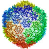
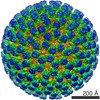
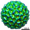
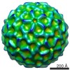
- EMDB-7317: P22 gp26minus asymmetric reconstruction C12 symmetry along Z-axis -
+ Open data

- Basic information
| Entry | Database: EMDB / ID: EMD-7317 | |||||||||
|---|---|---|---|---|---|---|---|---|---|---|
| Title | P22 gp26minus asymmetric reconstruction C12 symmetry along Z-axis | |||||||||
 Map data Map data | Bacteriophage P22 asymmetric reconstruction 12-fold symmetry along Z-axis | |||||||||
 Sample Sample |
| |||||||||
| Biological species |  Enterobacteria phage P22 (virus) Enterobacteria phage P22 (virus) | |||||||||
| Method | single particle reconstruction / cryo EM / Resolution: 12.5 Å | |||||||||
 Authors Authors | McNulty R / Johnson JE | |||||||||
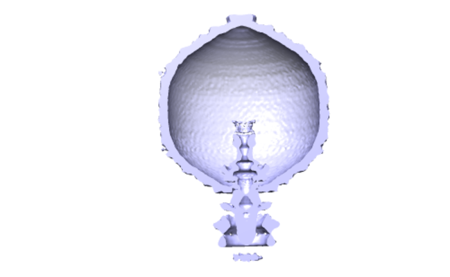
 Citation Citation | Journal: Biophys J / Year: 2018 Title: Cryo-EM Elucidation of the Structure of Bacteriophage P22 Virions after Genome Release. Authors: Reginald McNulty / Giovanni Cardone / Eddie B Gilcrease / Timothy S Baker / Sherwood R Casjens / John E Johnson /  Abstract: Genome ejection proteins are required to facilitate transport of bacteriophage P22 double-stranded DNA safely through membranes of Salmonella. The structures and locations of all proteins in the ...Genome ejection proteins are required to facilitate transport of bacteriophage P22 double-stranded DNA safely through membranes of Salmonella. The structures and locations of all proteins in the context of the mature virion are known, with the exception of three ejection proteins. Furthermore, the changes that occur to the proteins residing in the mature virion upon DNA release are not fully understood. We used cryogenic electron microscopy to obtain what is, to our knowledge, the first asymmetric reconstruction of mature bacteriophage P22 after double-stranded DNA has been extruded from the capsid-a state representative of one step during viral infection. Results of icosahedral and asymmetric reconstructions at estimated resolutions of 7.8 and 12.5 Å resolutions, respectively, are presented. The reconstruction shows tube-like protein density extending from the center of the tail assembly. The portal protein does not revert to the more contracted, procapsid state, but instead maintains an extended and splayed barrel structure. These structural details contribute to our understanding of the molecular mechanism of P22 phage infection and also set the foundation for future exploitation serving engineering purposes. | |||||||||
| History |
|
- Structure visualization
| Movie |
 Movie viewer Movie viewer |
|---|---|
| Structure viewer | EM map: SurfViewMolmilJmol/JSmol |
| Supplemental images |
- Downloads & links
-EMDB archive
| Map data | emd_7317.map.gz | 1.1 GB | EMDB map data format | |
|---|---|---|---|---|
| Header (meta data) | emd-7317-v30.xmlemd-7317.xml | 11.6 KB 11.6 KB | Display Display | EMDB header |
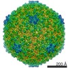
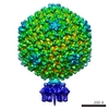
| Images |  emd_7317.png emd_7317.png | 72.1 KB | ||
| Archive directory |  http://ftp.pdbj.org/pub/emdb/structures/EMD-7317ftp://ftp.pdbj.org/pub/emdb/structures/EMD-7317 http://ftp.pdbj.org/pub/emdb/structures/EMD-7317ftp://ftp.pdbj.org/pub/emdb/structures/EMD-7317 | HTTPS FTP |
-Validation report
| Summary document | emd_7317_validation.pdf.gz | 78.1 KB | Display | EMDB validaton report |
|---|---|---|---|---|
| Full document | emd_7317_full_validation.pdf.gz | 77.1 KB | Display | |
| Data in XML | emd_7317_validation.xml.gz | 494 B | Display | |
| Arichive directory | https://ftp.pdbj.org/pub/emdb/validation_reports/EMD-7317ftp://ftp.pdbj.org/pub/emdb/validation_reports/EMD-7317 | HTTPS FTP |
-Related structure data

-Links
| EMDB pages | EMDB (EBI/PDBe) / EMDataResource |
|---|
-Map
| File | Download / File: emd_7317.map.gz / Format: CCP4 / Size: 1.9 GB / Type: IMAGE STORED AS FLOATING POINT NUMBER (4 BYTES) | ||||||||||||||||||||||||||||||||||||||||||||||||||||||||||||||||||||
|---|---|---|---|---|---|---|---|---|---|---|---|---|---|---|---|---|---|---|---|---|---|---|---|---|---|---|---|---|---|---|---|---|---|---|---|---|---|---|---|---|---|---|---|---|---|---|---|---|---|---|---|---|---|---|---|---|---|---|---|---|---|---|---|---|---|---|---|---|---|
| Annotation | Bacteriophage P22 asymmetric reconstruction 12-fold symmetry along Z-axis | ||||||||||||||||||||||||||||||||||||||||||||||||||||||||||||||||||||
| Projections & slices | Image control
Images are generated by Spider. | ||||||||||||||||||||||||||||||||||||||||||||||||||||||||||||||||||||
| Voxel size | X=Y=Z: 1.58 Å | ||||||||||||||||||||||||||||||||||||||||||||||||||||||||||||||||||||
| Density |
| ||||||||||||||||||||||||||||||||||||||||||||||||||||||||||||||||||||
| Symmetry | Space group: 1 | ||||||||||||||||||||||||||||||||||||||||||||||||||||||||||||||||||||
| Details | EMDB XML:
CCP4 map header:
| ||||||||||||||||||||||||||||||||||||||||||||||||||||||||||||||||||||
 Z (Sec.)
 Y (Row.)
 X (Col.)


-Supplemental data
- Sample components
-Entire : Enterobacteria phage P22
| Entire | Name: Enterobacteria phage P22 (virus) |
|---|---|
| Components |
|
-Supramolecule #1: Enterobacteria phage P22
| Supramolecule | Name: Enterobacteria phage P22 / type: virus / ID: 1 / Parent: 0 / NCBI-ID: 10754 / Sci species name: Enterobacteria phage P22 / Virus type: VIRION / Virus isolate: STRAIN / Virus enveloped: No / Virus empty: Yes |
|---|---|
| Host system | Organism:  Salmonella enterica (bacteria) Salmonella enterica (bacteria) |
-Experimental details
-Structure determination
| Method | cryo EM |
|---|---|
 Processing Processing | single particle reconstruction |
| Aggregation state | particle |
-Sample preparation
| Buffer | pH: 7.4 |
|---|---|
| Vitrification | Cryogen name: ETHANE |
- Electron microscopy
| Microscope | FEI TALOS ARCTICA |
|---|---|
| Image recording | Film or detector model: FEI FALCON II (4k x 4k) / Detector mode: COUNTING / Average electron dose: 57.0 e/Å2 |
| Electron beam | Acceleration voltage: 200 kV / Electron source:  FIELD EMISSION GUN FIELD EMISSION GUN |
| Electron optics | Illumination mode: FLOOD BEAM / Imaging mode: BRIGHT FIELD |
| Experimental equipment |  Model: Talos Arctica / Image courtesy: FEI Company |