Movie
Movie Controller


+ Open data

- Basic information
| Entry |  | ||||||||||||
|---|---|---|---|---|---|---|---|---|---|---|---|---|---|
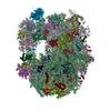
| Title | Structure of tuco-tuco ribosome (rotated, tRNAs, and mRNA) | ||||||||||||
 Map data Map data | |||||||||||||
 Sample Sample |
| ||||||||||||
 Keywords Keywords | Tuco tuco Ribosome / RIBOSOME | ||||||||||||
| Biological species |  Ctenomyidae (tuco-tucos) Ctenomyidae (tuco-tucos) | ||||||||||||
| Method | single particle reconstruction / cryo EM / Resolution: 3.3 Å | ||||||||||||
 Authors Authors | Gutierrez-Vargas C / De S / Maji S / Liu Z / Nieb M / Seluanov A / Gorbunova V / Frank J | ||||||||||||
| Funding support |  United States, 3 items United States, 3 items
| ||||||||||||
 Citation Citation | Journal: Nucleic Acids Res / Year: 2026 Title: Structures of naked mole-rat, tuco-tuco, and guinea pig ribosomes-is rRNA fragmentation linked to translational fidelity? Authors: Cristina Gutierrez-Vargas / Swastik De / Suvrajit Maji / Zheng Liu / Zhonghe Ke / Martina Nieß / Andrei Seluanov / Vera Gorbunova / Joachim Frank /   Abstract: Ribosomes are central to protein synthesis in all organisms. In mammals, the ribosome functional core is highly conserved. Remarkably, two rodent species, the naked mole-rat (NMR) and tuco-tuco, ...Ribosomes are central to protein synthesis in all organisms. In mammals, the ribosome functional core is highly conserved. Remarkably, two rodent species, the naked mole-rat (NMR) and tuco-tuco, display fragmented 28S ribosomal RNA (rRNA), coupled with high translational fidelity and long lifespan. The unusual ribosomal architecture in the NMR and tuco-tuco has been speculated to be linked to high translational fidelity. Here, we show, by single-particle cryo-electron microscopy, that despite the fragmentation of their rRNA, NMR and tuco-tuco ribosomes retain their core functional architecture. Compared to ribosomes of the guinea pig, a phylogenetically related rodent without 28S rRNA fragmentation, ribosomes of NMR and tuco-tuco exhibit poorly resolved density for certain expansion segments. In contrast, the structure of the guinea pig ribosome shows high similarity to the human ribosome. Enhanced translational fidelity in the NMR and tuco-tuco may stem from subtle, allosteric effects in dynamics, linked to rRNA fragmentation. | ||||||||||||
| History |
|
- Structure visualization
| Supplemental images |
|---|

- Downloads & links
-EMDB archive
| Map data | emd_72482.map.gz | 193.4 MB |  EMDB map data format EMDB map data format | |
|---|---|---|---|---|
| Header (meta data) | emd-72482-v30.xmlemd-72482.xml | 97.4 KB 97.4 KB | Display Display | EMDB header |
| FSC (resolution estimation) | emd_72482_fsc.xml | 18.3 KB | Display | FSC data file |
| Images |  emd_72482.png emd_72482.png | 148.6 KB | ||
| Filedesc metadata | emd-72482.cif.gz | 19.7 KB | ||
| Others | emd_72482_half_map_1.map.gzemd_72482_half_map_2.map.gz | 194 MB 194 MB | ||
| Archive directory |  http://ftp.pdbj.org/pub/emdb/structures/EMD-72482ftp://ftp.pdbj.org/pub/emdb/structures/EMD-72482 http://ftp.pdbj.org/pub/emdb/structures/EMD-72482ftp://ftp.pdbj.org/pub/emdb/structures/EMD-72482 | HTTPS FTP |
-Related structure data
| Related structure data |  9y4gMC  9y42C  9y44C  9y49C  9y4hC  9zrgC M: atomic model generated by this map C: citing same article ( |
|---|


-Links
| EMDB pages | EMDB (EBI/PDBe) / EMDataResource |
|---|
-Map
| File | Download / File: emd_72482.map.gz / Format: CCP4 / Size: 244.1 MB / Type: IMAGE STORED AS FLOATING POINT NUMBER (4 BYTES) | ||||||||||||||||||||||||||||||||||||
|---|---|---|---|---|---|---|---|---|---|---|---|---|---|---|---|---|---|---|---|---|---|---|---|---|---|---|---|---|---|---|---|---|---|---|---|---|---|
| Projections & slices | Image control
Images are generated by Spider. | ||||||||||||||||||||||||||||||||||||
| Voxel size | X=Y=Z: 1.045 Å | ||||||||||||||||||||||||||||||||||||
| Density |
| ||||||||||||||||||||||||||||||||||||
| Symmetry | Space group: 1 | ||||||||||||||||||||||||||||||||||||
| Details | EMDB XML:
|
 Z (Sec.)
 Y (Row.)
 X (Col.)


-Supplemental data
-Half map: #2
| File | emd_72482_half_map_1.map | ||||||||||||
|---|---|---|---|---|---|---|---|---|---|---|---|---|---|
| Projections & Slices |
| ||||||||||||
| Density Histograms |


-Half map: #1
| File | emd_72482_half_map_2.map | ||||||||||||
|---|---|---|---|---|---|---|---|---|---|---|---|---|---|
| Projections & Slices |
| ||||||||||||
| Density Histograms |


- Sample components
+Entire : Tuco tuco Ribosome
+Supramolecule #1: Tuco tuco Ribosome
+Macromolecule #1: Guanine nucleotide-binding protein subunit beta-2-like 1
+Macromolecule #2: 40S ribosomal protein S20
+Macromolecule #3: 40S ribosomal protein S10
+Macromolecule #4: 40S ribosomal protein S12
+Macromolecule #5: 40S ribosomal protein S18
+Macromolecule #6: 40S ribosomal protein S29
+Macromolecule #7: 40S ribosomal protein S17
+Macromolecule #8: 40S ribosomal protein S15
+Macromolecule #9: 40S ribosomal protein S19
+Macromolecule #10: 40S ribosomal protein S25
+Macromolecule #11: 40S ribosomal protein S28
+Macromolecule #12: 40S ribosomal protein S3
+Macromolecule #13: 40S ribosomal protein S27a
+Macromolecule #14: 40S ribosomal protein S5
+Macromolecule #15: 40S ribosomal protein S16
+Macromolecule #17: 40S ribosomal protein S14
+Macromolecule #18: 40S ribosomal protein S23
+Macromolecule #19: 40S ribosomal protein S13
+Macromolecule #20: 40S ribosomal protein S11
+Macromolecule #21: 40S ribosomal protein S3a
+Macromolecule #22: 40S ribosomal protein SA
+Macromolecule #23: 40S ribosomal protein S21
+Macromolecule #24: 40S ribosomal protein S24
+Macromolecule #25: 40S ribosomal protein S26
+Macromolecule #26: 40S ribosomal protein S27
+Macromolecule #27: 40S ribosomal protein S30
+Macromolecule #28: 40S ribosomal protein S9
+Macromolecule #29: 40S ribosomal protein S4, X isoform
+Macromolecule #30: 40S ribosomal protein S2
+Macromolecule #31: 40S ribosomal protein S6
+Macromolecule #32: 40S ribosomal protein S7
+Macromolecule #33: 40S ribosomal protein S15a
+Macromolecule #34: 40S ribosomal protein S8
+Macromolecule #35: 60S ribosomal protein L19
+Macromolecule #36: 60S ribosomal protein L24
+Macromolecule #37: 60S ribosomal protein L10a
+Macromolecule #38: 60S ribosomal protein L13a
+Macromolecule #39: 60S ribosomal protein L13
+Macromolecule #40: 60S ribosomal protein L23
+Macromolecule #41: 60S ribosomal protein L14
+Macromolecule #42: 60S ribosomal protein L27a
+Macromolecule #43: 60S ribosomal protein L15
+Macromolecule #44: 60S ribosomal protein L10-like
+Macromolecule #45: 60S ribosomal protein L5
+Macromolecule #46: 60S ribosomal protein L18
+Macromolecule #47: 60S ribosomal protein L8
+Macromolecule #48: 60S ribosomal protein L18a
+Macromolecule #49: 60S ribosomal protein L21
+Macromolecule #50: 60S ribosomal protein L17
+Macromolecule #51: 60S ribosomal protein L22
+Macromolecule #52: 60S ribosomal protein L23a
+Macromolecule #53: 60S ribosomal protein L26
+Macromolecule #54: 60S ribosomal protein L27
+Macromolecule #55: 60S ribosomal protein L28
+Macromolecule #56: 60S ribosomal protein L35
+Macromolecule #57: 60S ribosomal protein L29
+Macromolecule #58: 60S ribosomal protein L3
+Macromolecule #59: 60S ribosomal protein L7
+Macromolecule #60: 60S ribosomal protein L31
+Macromolecule #61: 60S ribosomal protein L32
+Macromolecule #62: 60S ribosomal protein L35a
+Macromolecule #63: 60S ribosomal protein L34
+Macromolecule #64: 60S ribosomal protein L36
+Macromolecule #65: 60S ribosomal protein L37
+Macromolecule #66: 60S ribosomal protein L38
+Macromolecule #67: 60S ribosomal protein L39
+Macromolecule #68: 60S ribosomal protein L4
+Macromolecule #69: 60S ribosomal protein L40
+Macromolecule #70: 60S ribosomal protein L41
+Macromolecule #71: 60S ribosomal protein L37a
+Macromolecule #72: 60S ribosomal protein L36a
+Macromolecule #73: 60S ribosomal protein L11
+Macromolecule #74: 60S ribosomal protein L9
+Macromolecule #75: 60S ribosomal protein L6
+Macromolecule #76: 60S ribosomal protein L7a
+Macromolecule #16: 18S ribosomal RNA
+Macromolecule #77: LSU-alpha rRNA
+Macromolecule #78: 5S ribosomal RNA
+Macromolecule #79: 5.8S rRNA (157-MER)
+Macromolecule #80: tRNA (78-MER)
+Macromolecule #81: tRNA (77-MER)
+Macromolecule #82: RNA (5'-R(P*UP*UP*UP*UP*UP*UP*UP*UP*UP*UP*UP*UP*U*(MG))-3')
+Macromolecule #83: LSU-beta rRNA (2108-MER)
+Macromolecule #84: MAGNESIUM ION
+Macromolecule #85: water

-Experimental details
-Structure determination
| Method | cryo EM |
|---|---|
 Processing Processing | single particle reconstruction |
| Aggregation state | particle |
-Sample preparation
| Buffer | pH: 7.5 Component:
| |||||||||||||||
|---|---|---|---|---|---|---|---|---|---|---|---|---|---|---|---|---|
| Grid | Model: Quantifoil R2/2 / Material: COPPER / Support film - Material: CARBON / Support film - topology: HOLEY / Pretreatment - Type: GLOW DISCHARGE | |||||||||||||||
| Vitrification | Cryogen name: ETHANE-PROPANE |
- Electron microscopy
| Microscope | TFS KRIOS |
|---|---|
| Software | Name: EPU |
| Image recording | Film or detector model: FEI FALCON II (4k x 4k) / Detector mode: INTEGRATING / Number real images: 17093 / Average electron dose: 30.0 e/Å2 |
| Electron beam | Acceleration voltage: 300 kV / Electron source:  FIELD EMISSION GUN FIELD EMISSION GUN |
| Electron optics | Illumination mode: FLOOD BEAM / Imaging mode: BRIGHT FIELD / Nominal defocus max: 3.0 µm / Nominal defocus min: 1.0 µm |
| Experimental equipment |  Model: Titan Krios / Image courtesy: FEI Company |