 Movie
Movie Controller


+ Open data

- Basic information
| Entry | Database: EMDB / ID: EMD-7131 | |||||||||
|---|---|---|---|---|---|---|---|---|---|---|
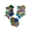
| Title | Chromatin-modifying complex | |||||||||
 Map data Map data | Structure of the Saccharomyces cerevisiae NuA4 histone acetyltransferase complex | |||||||||
 Sample Sample |
| |||||||||
| Biological species |  | |||||||||
| Method | single particle reconstruction / negative staining / Resolution: 26.5 Å | |||||||||
 Authors Authors | Setiaputra DT / Dalwadi U / Yip CK | |||||||||
 Citation Citation | Journal: Mol Cell Biol / Year: 2018 Title: Molecular Architecture of the Essential Yeast Histone Acetyltransferase Complex NuA4 Redefines Its Multimodularity. Authors: Dheva Setiaputra / Salar Ahmad / Udit Dalwadi / Anne-Lise Steunou / Shan Lu / James D Ross / Meng-Qiu Dong / Jacques Côté / Calvin K Yip /   Abstract: Conserved from yeast to humans, the NuA4 histone acetyltransferase is a large multisubunit complex essential for cell viability through the regulation of gene expression, genome maintenance, ...Conserved from yeast to humans, the NuA4 histone acetyltransferase is a large multisubunit complex essential for cell viability through the regulation of gene expression, genome maintenance, metabolism, and cell fate during development and stress. How the different NuA4 subunits work in concert with one another to perform these diverse functions remains unclear, and addressing this central question requires a comprehensive understanding of NuA4's molecular architecture and subunit organization. We have determined the structure of fully assembled native yeast NuA4 by single-particle electron microscopy. Our data revealed that NuA4 adopts a trilobal overall architecture, with each of the three lobes constituted by one or two functional modules. By performing cross-linking coupled to mass spectrometry analysis and protein interaction studies, we further mapped novel intermolecular interfaces within NuA4. Finally, we combined these new data with other known structural information of NuA4 subunits and subassemblies to construct a multiscale model to illustrate how the different NuA4 subunits and modules are spatially arranged. This model shows that the multiple chromatin reader domains are clustered together around the catalytic core, suggesting that NuA4's multimodular architecture enables it to engage in multivalent interactions with its nucleosome substrate. | |||||||||
| History |
|
- Structure visualization
| Movie |
 Movie viewer Movie viewer |
|---|---|
| Structure viewer | EM map: SurfViewMolmilJmol/JSmol |
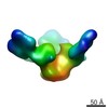
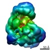
| Supplemental images |
 UCSF Chimera
- Downloads & links
-EMDB archive
| Map data | emd_7131.map.gz | 1 MB | EMDB map data format | |
|---|---|---|---|---|
| Header (meta data) | emd-7131-v30.xmlemd-7131.xml | 14.6 KB 14.6 KB | Display Display | EMDB header |
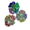
| Images | 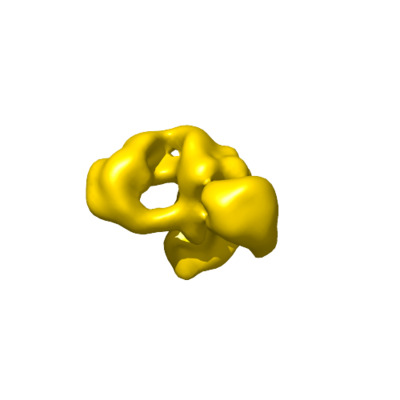 emd_7131.png emd_7131.png | 32.4 KB | ||
| Archive directory |  http://ftp.pdbj.org/pub/emdb/structures/EMD-7131ftp://ftp.pdbj.org/pub/emdb/structures/EMD-7131 http://ftp.pdbj.org/pub/emdb/structures/EMD-7131ftp://ftp.pdbj.org/pub/emdb/structures/EMD-7131 | HTTPS FTP |
-Related structure data
| Similar structure data |
|---|
-Links
| EMDB pages | EMDB (EBI/PDBe) / EMDataResource |
|---|
-Map
| File | Download / File: emd_7131.map.gz / Format: CCP4 / Size: 1.4 MB / Type: IMAGE STORED AS FLOATING POINT NUMBER (4 BYTES) | ||||||||||||||||||||||||||||||||||||||||||||||||||||||||||||||||||||
|---|---|---|---|---|---|---|---|---|---|---|---|---|---|---|---|---|---|---|---|---|---|---|---|---|---|---|---|---|---|---|---|---|---|---|---|---|---|---|---|---|---|---|---|---|---|---|---|---|---|---|---|---|---|---|---|---|---|---|---|---|---|---|---|---|---|---|---|---|---|
| Annotation | Structure of the Saccharomyces cerevisiae NuA4 histone acetyltransferase complex | ||||||||||||||||||||||||||||||||||||||||||||||||||||||||||||||||||||
| Projections & slices | Image control
Images are generated by Spider. | ||||||||||||||||||||||||||||||||||||||||||||||||||||||||||||||||||||
| Voxel size | X=Y=Z: 7 Å | ||||||||||||||||||||||||||||||||||||||||||||||||||||||||||||||||||||
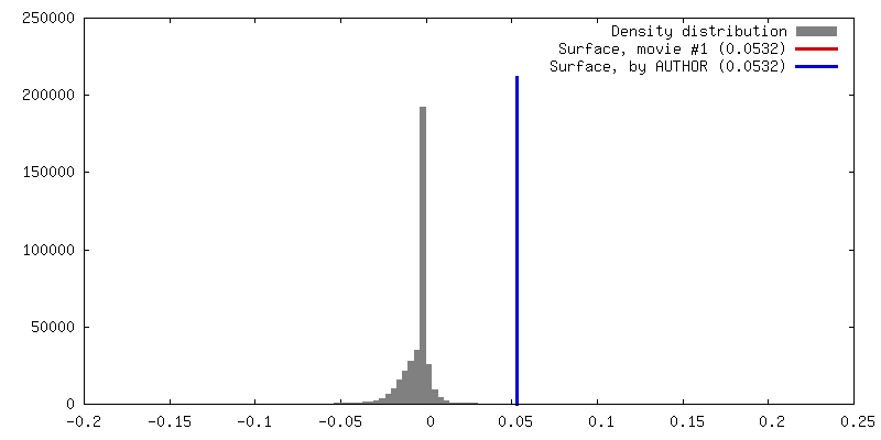
| Density |
| ||||||||||||||||||||||||||||||||||||||||||||||||||||||||||||||||||||
| Symmetry | Space group: 1 | ||||||||||||||||||||||||||||||||||||||||||||||||||||||||||||||||||||
| Details | EMDB XML:
CCP4 map header:
| ||||||||||||||||||||||||||||||||||||||||||||||||||||||||||||||||||||
 Z (Sec.)
 Y (Row.)
 X (Col.)


-Supplemental data
- Sample components
-Entire : Chromatin-modifying complex
| Entire | Name: Chromatin-modifying complex |
|---|---|
| Components |
|
-Supramolecule #1: Chromatin-modifying complex
| Supramolecule | Name: Chromatin-modifying complex / type: complex / ID: 1 / Parent: 0 |
|---|---|
| Source (natural) | Organism: |
| Molecular weight | Theoretical: 1 MDa |
-Experimental details
-Structure determination
| Method | negative staining |
|---|---|
 Processing Processing | single particle reconstruction |
| Aggregation state | particle |
-Sample preparation
| Buffer | pH: 7.4 Component:
| |||||||||||||||||||||
|---|---|---|---|---|---|---|---|---|---|---|---|---|---|---|---|---|---|---|---|---|---|---|
| Staining | Type: NEGATIVE / Material: Uranyl formate Details: Negatively-stained EM specimens were prepared by uranyl formate staining on continuous carbon-overlaid copper grids. | |||||||||||||||||||||
| Grid | Model: Ted Pella / Material: COPPER / Mesh: 400 / Support film - Material: FORMVAR / Support film - topology: CONTINUOUS / Pretreatment - Type: GLOW DISCHARGE / Pretreatment - Atmosphere: AIR / Pretreatment - Pressure: 101.325 kPa | |||||||||||||||||||||
| Details | Complexes were purified from yeast via affinity purification for a FLAG-tagged subunit. Purified complexes were subjected to glycerol gradient ultracentrifugation, fractionated, and analyzed by negative-stain electron microscopy. |
- Electron microscopy
| Microscope | FEI TECNAI SPIRIT |
|---|---|
| Image recording | Film or detector model: FEI EAGLE (4k x 4k) / Digitization - Dimensions - Width: 1365 pixel / Digitization - Dimensions - Height: 1365 pixel / Number grids imaged: 1 / Number real images: 251 / Average exposure time: 1.0 sec. / Average electron dose: 25.0 e/Å2 |
| Electron beam | Acceleration voltage: 120 kV / Electron source: LAB6 |
| Electron optics | Illumination mode: FLOOD BEAM / Imaging mode: BRIGHT FIELD / Nominal defocus max: 1.5 µm / Nominal defocus min: 1.0 µm / Nominal magnification: 49000 |
| Sample stage | Specimen holder model: SIDE ENTRY, EUCENTRIC / Cooling holder cryogen: NITROGEN |
| Experimental equipment |  Model: Tecnai Spirit / Image courtesy: FEI Company |
