 Movie
Movie Controller


+ Open data

- Basic information
| Entry |  | |||||||||||||||
|---|---|---|---|---|---|---|---|---|---|---|---|---|---|---|---|---|
| Title | S. aureus YhaM D193A hexamer, D3 refinement | |||||||||||||||
 Map data Map data | Sharpened Homogeneous Refinement map, used for modeling. Bsharpen = 126.9 | |||||||||||||||
 Sample Sample |
| |||||||||||||||
 Keywords Keywords | exonuclease / translation / RNA / RNA BINDING PROTEIN | |||||||||||||||
| Function / homology |  Function and homology information Function and homology information | |||||||||||||||
| Biological species |  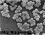 Staphylococcus aureus (bacteria) Staphylococcus aureus (bacteria) | |||||||||||||||
| Method | single particle reconstruction / cryo EM / Resolution: 2.36 Å | |||||||||||||||
 Authors Authors | Mattingly JM / Tanquary JR / Dunham CM | |||||||||||||||
| Funding support |  United States, 4 items United States, 4 items
| |||||||||||||||
 Citation Citation | Journal: Proc Natl Acad Sci U S A / Year: 2026 Title: Structural insights into RNA recognition by the exoribonuclease YhaM. Authors: Jacob M Mattingly / Anna Lipońska / Julia Tanquary / Mee-Ngan F Yap / Christine M Dunham / Abstract: Bacterial ribonucleases regulate gene expression in response to environmental stress and host interactions. In the hibernation-promoting factor (Hpf) induces the formation of RNase R-resistant 100S ...Bacterial ribonucleases regulate gene expression in response to environmental stress and host interactions. In the hibernation-promoting factor (Hpf) induces the formation of RNase R-resistant 100S ribosomes. We previously showed that the 3'-5' exoribonuclease YhaM cleaves the transcript, reducing Hpf synthesis and leading to ribosome degradation. No structure of any YhaM homolog bound to RNA is available, and biological investigations of YhaM remain limited. Here, we find that deletion of attenuates virulence in a infection model. We further determined electron cryomicroscopy structures of YhaM-RNA complexes. YhaM adopts a hexameric complex arranged in a ring, with its N-terminal oligonucleotide/oligosaccharide-binding (OB) domains positioned on both sides of the ring while the catalytic histidine/aspartate-rich (HD) domain active sites are buried within the interior. The OB-1'' domains recognize the hairpin by the formation of complementary minor groove interactions. RNA binding by two YhaM OB domains is mediated through engagement of both the backbones and nucleobases of the RNA substrate, where stacking of aromatic residues and nucleobases likely contributes to substrate recognition. Structures of YhaM bound to a single-stranded RNA reveal how the 3' ends of two RNAs are positioned within the HD domain poised for catalysis. Although six YhaM active sites are present, only two engage in RNA cleavage and further point to the importance of the remaining YhaM monomers as structural scaffolds for guiding RNA to the active site. In summary, these findings provide insights into the unique assembly of an understudied bacterial RNase. | |||||||||||||||
| History |
|
- Structure visualization
| Supplemental images |
|---|

- Downloads & links
-EMDB archive
| Map data | emd_70886.map.gz | 59.8 MB | EMDB map data format | |
|---|---|---|---|---|
| Header (meta data) | emd-70886-v30.xmlemd-70886.xml | 30.2 KB 30.2 KB | Display Display | EMDB header |
| FSC (resolution estimation) | emd_70886_fsc.xml | 8.4 KB | Display | FSC data file |
| Images |  emd_70886.png emd_70886.png | 166.8 KB | ||
| Masks | emd_70886_msk_1.map | 64 MB | Mask map | |
| Filedesc metadata | emd-70886.cif.gz | 7.5 KB | ||
| Others | emd_70886_additional_1.map.gzemd_70886_half_map_1.map.gzemd_70886_half_map_2.map.gz | 31.7 MB 59.3 MB 59.3 MB | ||
| Archive directory |  https://data.pdbj.org/pub/emdb/structures/EMD-70886ftp://data.pdbj.org/pub/emdb/structures/EMD-70886 https://data.pdbj.org/pub/emdb/structures/EMD-70886ftp://data.pdbj.org/pub/emdb/structures/EMD-70886 | HTTPS FTP |
-Related structure data
| Related structure data |  9ov1MC 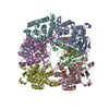 9o7tC  9ypeC  9ypfC C: citing same article ( M: atomic model generated by this map |
|---|---|
| Similar structure data |


-Links
| EMDB pages | EMDB (EBI/PDBe) / EMDataResource |
|---|---|
| Related items in Molecule of the Month |

-Map
| File | Download / File: emd_70886.map.gz / Format: CCP4 / Size: 64 MB / Type: IMAGE STORED AS FLOATING POINT NUMBER (4 BYTES) | ||||||||||||||||||||||||||||||||||||
|---|---|---|---|---|---|---|---|---|---|---|---|---|---|---|---|---|---|---|---|---|---|---|---|---|---|---|---|---|---|---|---|---|---|---|---|---|---|
| Annotation | Sharpened Homogeneous Refinement map, used for modeling. Bsharpen = 126.9 | ||||||||||||||||||||||||||||||||||||
| Projections & slices | Image control
Images are generated by Spider. | ||||||||||||||||||||||||||||||||||||
| Voxel size | X=Y=Z: 0.825 Å | ||||||||||||||||||||||||||||||||||||
| Density |
| ||||||||||||||||||||||||||||||||||||
| Symmetry | Space group: 1 | ||||||||||||||||||||||||||||||||||||
| Details | EMDB XML:
|
 Z (Sec.)
 Y (Row.)
 X (Col.)


-Supplemental data
-Mask #1
| File | emd_70886_msk_1.map | ||||||||||||
|---|---|---|---|---|---|---|---|---|---|---|---|---|---|
| Projections & Slices |
| ||||||||||||
| Density Histograms |


-Additional map: Unsharpened map from Homogeneous Refinement
| File | emd_70886_additional_1.map | ||||||||||||
|---|---|---|---|---|---|---|---|---|---|---|---|---|---|
| Annotation | Unsharpened map from Homogeneous Refinement | ||||||||||||
| Projections & Slices |
| ||||||||||||
| Density Histograms |


-Half map: Half-map A from Homogeneous Refinement
| File | emd_70886_half_map_1.map | ||||||||||||
|---|---|---|---|---|---|---|---|---|---|---|---|---|---|
| Annotation | Half-map A from Homogeneous Refinement | ||||||||||||
| Projections & Slices |
| ||||||||||||
| Density Histograms |


-Half map: Half-map B from Homogeneous Refinement
| File | emd_70886_half_map_2.map | ||||||||||||
|---|---|---|---|---|---|---|---|---|---|---|---|---|---|
| Annotation | Half-map B from Homogeneous Refinement | ||||||||||||
| Projections & Slices |
| ||||||||||||
| Density Histograms |


- Sample components
-Entire : YhaM D193A hexamer
| Entire | Name: YhaM D193A hexamer |
|---|---|
| Components |
|
-Supramolecule #1: YhaM D193A hexamer
| Supramolecule | Name: YhaM D193A hexamer / type: complex / ID: 1 / Parent: 0 / Macromolecule list: #1 |
|---|---|
| Source (natural) | Organism: Staphylococcus aureus (bacteria) |
-Macromolecule #1: YhaM
| Macromolecule | Name: YhaM / type: protein_or_peptide / ID: 1 / Number of copies: 6 / Enantiomer: LEVO |
|---|---|
| Source (natural) | Organism: Staphylococcus aureus (bacteria) |
| Molecular weight | Theoretical: 35.785797 KDa |
| Recombinant expression | Organism:  Escherichia coli BL21(DE3) (bacteria) Escherichia coli BL21(DE3) (bacteria) |
| Sequence | String: MRNIENLNPG DSVDHFFLVH KATQGVTAQG KDYMTLHLQD KSGEIEAKFW TATKNDMATI KPEEIVHVKG DIINYRGNKQ MKVNQIRLA TTEDQLKTEQ FVDGAPLSPA EIQEEISHYL LDIENANLQR ITRHLLKKYQ ERFYTYPAAS SHHHNFASGL S YHVLTMLR ...String: MRNIENLNPG DSVDHFFLVH KATQGVTAQG KDYMTLHLQD KSGEIEAKFW TATKNDMATI KPEEIVHVKG DIINYRGNKQ MKVNQIRLA TTEDQLKTEQ FVDGAPLSPA EIQEEISHYL LDIENANLQR ITRHLLKKYQ ERFYTYPAAS SHHHNFASGL S YHVLTMLR IAKSICDIYP LLNKSLLYSG IILHAIGKVR ELSGPVATSY TVEGNLLGHI SIASDEVVEA ARELNIEGEE IM LLRHMIL SHHGKLEYGS PKLPYLKEAE ILCYIDNIDA RMNMFEKAYK KTDKGQFTDK IFGLENRRFY NPESLD UniProtKB: Cmp-binding-factor 1 |
-Macromolecule #2: MAGNESIUM ION
| Macromolecule | Name: MAGNESIUM ION / type: ligand / ID: 2 / Number of copies: 12 / Formula: MG |
|---|---|
| Molecular weight | Theoretical: 24.305 Da |
-Macromolecule #3: PHOSPHATE ION
| Macromolecule | Name: PHOSPHATE ION / type: ligand / ID: 3 / Number of copies: 6 / Formula: PO4 |
|---|---|
| Molecular weight | Theoretical: 94.971 Da |
| Chemical component information | 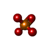 ChemComp-PO4: |
-Experimental details
-Structure determination
| Method | cryo EM |
|---|---|
 Processing Processing | single particle reconstruction |
| Aggregation state | particle |
-Sample preparation
| Concentration | 1.1 mg/mL | |||||||||||||||
|---|---|---|---|---|---|---|---|---|---|---|---|---|---|---|---|---|
| Buffer | pH: 8 Component:
| |||||||||||||||
| Grid | Model: C-flat-1.2/1.3 / Material: GOLD / Mesh: 300 / Support film - Material: CARBON / Support film - topology: HOLEY / Pretreatment - Type: PLASMA CLEANING / Pretreatment - Time: 15 sec. / Pretreatment - Atmosphere: OTHER | |||||||||||||||
| Vitrification | Cryogen name: ETHANE / Chamber humidity: 100 % / Chamber temperature: 277 K / Instrument: FEI VITROBOT MARK IV |
- Electron microscopy
| Microscope | TFS KRIOS |
|---|---|
| Specialist optics | Energy filter - Name: GIF Bioquantum / Energy filter - Slit width: 20 eV |
| Image recording | Film or detector model: GATAN K3 BIOQUANTUM (6k x 4k) / Digitization - Dimensions - Width: 5760 pixel / Digitization - Dimensions - Height: 4092 pixel / Number grids imaged: 1 / Number real images: 14007 / Average exposure time: 1.6 sec. / Average electron dose: 47.0 e/Å2 |
| Electron beam | Acceleration voltage: 300 kV / Electron source:  FIELD EMISSION GUN FIELD EMISSION GUN |
| Electron optics | Illumination mode: FLOOD BEAM / Imaging mode: BRIGHT FIELD / Cs: 2.7 mm / Nominal defocus max: 2.0 µm / Nominal defocus min: 0.5 µm / Nominal magnification: 105000 |
| Sample stage | Specimen holder model: FEI TITAN KRIOS AUTOGRID HOLDER / Cooling holder cryogen: NITROGEN |
| Experimental equipment |  Model: Titan Krios / Image courtesy: FEI Company |
+Image processing

-Atomic model buiding 1
| Initial model | Chain - Source name: Other / Chain - Initial model type: other / Details: Initial model built de novo using ModelAngelo |
|---|---|
| Details | Model was built into main EM map de novo using ModelAngelo, then flexibly fit using PHENIX real-space model refinement. |
| Refinement | Space: REAL / Protocol: FLEXIBLE FIT / Overall B value: 126.9 / Target criteria: Cross-correlation coefficient (CCmask) |
| Output model | PDB-9ov1: |
