 Movie
Movie Controller

[English] 日本語
 Yorodumi
- EMDB-7060: Focused map on the aCTD-CAP region of the class-I bacterial trans... -
+ Open data

- Basic information
| Entry | Database: EMDB / ID: EMD-7060 | |||||||||
|---|---|---|---|---|---|---|---|---|---|---|
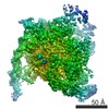
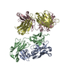
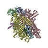
| Title | Focused map on the aCTD-CAP region of the class-I bacterial transcription activation complex | |||||||||
 Map data Map data | Focused map of class I transcription activation complex | |||||||||
 Sample Sample |
| |||||||||
| Biological species |  | |||||||||
| Method | single particle reconstruction / cryo EM / Resolution: 10.0 Å | |||||||||
 Authors Authors | Liu B / Hong C / Huang R / Yu Z / Steitz TA | |||||||||
| Funding support |  United States, 1 items United States, 1 items
| |||||||||
 Citation Citation | Journal: Science / Year: 2017 Title: Structural basis of bacterial transcription activation. Authors: Bin Liu / Chuan Hong / Rick K Huang / Zhiheng Yu / Thomas A Steitz / Abstract: In bacteria, the activation of gene transcription at many promoters is simple and only involves a single activator. The cyclic adenosine 3',5'-monophosphate receptor protein (CAP), a classic ...In bacteria, the activation of gene transcription at many promoters is simple and only involves a single activator. The cyclic adenosine 3',5'-monophosphate receptor protein (CAP), a classic activator, is able to activate transcription independently through two different mechanisms. Understanding the class I mechanism requires an intact transcription activation complex (TAC) structure at a high resolution. Here we report a high-resolution cryo-electron microscopy structure of an intact class I TAC containing a CAP dimer, a σ-RNA polymerase (RNAP) holoenzyme, a complete class I CAP-dependent promoter DNA, and a de novo synthesized RNA oligonucleotide. The structure shows how CAP wraps the upstream DNA and how the interactions recruit RNAP. Our study provides a structural basis for understanding how activators activate transcription through the class I recruitment mechanism. | |||||||||
| History |
|
- Structure visualization
| Movie |
 Movie viewer Movie viewer |
|---|---|
| Structure viewer | EM map: SurfViewMolmilJmol/JSmol |
| Supplemental images |

- Downloads & links
-EMDB archive
| Map data | emd_7060.map.gz | 615 KB | EMDB map data format | |
|---|---|---|---|---|
| Header (meta data) | emd-7060-v30.xmlemd-7060.xml | 13.6 KB 13.6 KB | Display Display | EMDB header |
| Images |  emd_7060.png emd_7060.png | 150.2 KB | ||
| Archive directory |  http://ftp.pdbj.org/pub/emdb/structures/EMD-7060ftp://ftp.pdbj.org/pub/emdb/structures/EMD-7060 http://ftp.pdbj.org/pub/emdb/structures/EMD-7060ftp://ftp.pdbj.org/pub/emdb/structures/EMD-7060 | HTTPS FTP |
-Related structure data


-Links
| EMDB pages | EMDB (EBI/PDBe) / EMDataResource |
|---|
-Map
| File | Download / File: emd_7060.map.gz / Format: CCP4 / Size: 30.5 MB / Type: IMAGE STORED AS FLOATING POINT NUMBER (4 BYTES) | ||||||||||||||||||||||||||||||||||||||||||||||||||||||||||||||||||||
|---|---|---|---|---|---|---|---|---|---|---|---|---|---|---|---|---|---|---|---|---|---|---|---|---|---|---|---|---|---|---|---|---|---|---|---|---|---|---|---|---|---|---|---|---|---|---|---|---|---|---|---|---|---|---|---|---|---|---|---|---|---|---|---|---|---|---|---|---|---|
| Annotation | Focused map of class I transcription activation complex | ||||||||||||||||||||||||||||||||||||||||||||||||||||||||||||||||||||
| Projections & slices | Image control
Images are generated by Spider. | ||||||||||||||||||||||||||||||||||||||||||||||||||||||||||||||||||||
| Voxel size | X=Y=Z: 1.35 Å | ||||||||||||||||||||||||||||||||||||||||||||||||||||||||||||||||||||
| Density |
| ||||||||||||||||||||||||||||||||||||||||||||||||||||||||||||||||||||
| Symmetry | Space group: 1 | ||||||||||||||||||||||||||||||||||||||||||||||||||||||||||||||||||||
| Details | EMDB XML:
CCP4 map header:
| ||||||||||||||||||||||||||||||||||||||||||||||||||||||||||||||||||||
 Z (Sec.)
 Y (Row.)
 X (Col.)


-Supplemental data
- Sample components
-Entire : The aCTD-CAP region of the class-I bacterial transcription activa...
| Entire | Name: The aCTD-CAP region of the class-I bacterial transcription activation complex |
|---|---|
| Components |
|
-Supramolecule #1: The aCTD-CAP region of the class-I bacterial transcription activa...
| Supramolecule | Name: The aCTD-CAP region of the class-I bacterial transcription activation complex type: complex / ID: 1 / Parent: 0 / Details: Focused map only on the aCTD-CAP region |
|---|---|
| Source (natural) | Organism: |
| Recombinant expression | Organism: |
| Molecular weight | Theoretical: 590 KDa |
-Experimental details
-Structure determination
| Method | cryo EM |
|---|---|
 Processing Processing | single particle reconstruction |
| Aggregation state | particle |
-Sample preparation
| Concentration | 3 mg/mL |
|---|---|
| Buffer | pH: 7.5 Details: 20 mM TRIS pH 7.5, 50 mM sodium chloride, 0.1mM EDTA, 5 mM MgCl2 |
| Grid | Model: Quantifoil R1.2/1.3 / Material: GOLD / Mesh: 400 / Pretreatment - Type: GLOW DISCHARGE / Pretreatment - Atmosphere: AIR / Pretreatment - Pressure: 0.038 kPa |
| Vitrification | Cryogen name: ETHANE / Chamber humidity: 100 % / Chamber temperature: 277 K / Instrument: FEI VITROBOT MARK IV / Details: 3 seconds blotting. |
- Electron microscopy
| Microscope | FEI TITAN KRIOS |
|---|---|
| Temperature | Min: 98.0 K / Max: 98.0 K |
| Specialist optics | Energy filter - Name: GIF Quantum LS / Energy filter - Lower energy threshold: 0 eV / Energy filter - Upper energy threshold: 20 eV |
| Details | Cs Corrector |
| Image recording | Film or detector model: GATAN K2 SUMMIT (4k x 4k) / Detector mode: SUPER-RESOLUTION / Digitization - Dimensions - Width: 7676 pixel / Digitization - Dimensions - Height: 7420 pixel / Digitization - Sampling interval: 5.0 µm / Digitization - Frames/image: 1-40 / Number grids imaged: 1 / Number real images: 2382 / Average exposure time: 0.25 sec. / Average electron dose: 1.37 e/Å2 |
| Electron beam | Acceleration voltage: 300 kV / Electron source:  FIELD EMISSION GUN FIELD EMISSION GUN |
| Electron optics | C2 aperture diameter: 70.0 µm / Calibrated defocus max: 2.6 µm / Calibrated defocus min: 1.2 µm / Calibrated magnification: 37037 / Illumination mode: FLOOD BEAM / Imaging mode: BRIGHT FIELD / Cs: 0.01 mm / Nominal defocus max: 2.5 µm / Nominal defocus min: 1.3 µm / Nominal magnification: 81000 |
| Sample stage | Specimen holder model: FEI TITAN KRIOS AUTOGRID HOLDER / Cooling holder cryogen: NITROGEN |
| Experimental equipment |  Model: Titan Krios / Image courtesy: FEI Company |
+Image processing
-Atomic model buiding 1
| Refinement | Space: REAL / Protocol: RIGID BODY FIT / Target criteria: correlation coefficient |
|---|
