 Movie
Movie Controller

[English] 日本語
 Yorodumi
- EMDB-70421: The structure of a Fungal Cyanide Hydratase from Gloeocercospora ... -
+ Open data

- Basic information
| Entry |  | ||||||||||||
|---|---|---|---|---|---|---|---|---|---|---|---|---|---|
| Title | The structure of a Fungal Cyanide Hydratase from Gloeocercospora sorghi | ||||||||||||
 Map data Map data | The structure of a Fungal Cyanide Hydratase from Gloeocercospora sorghi | ||||||||||||
 Sample Sample |
| ||||||||||||
 Keywords Keywords | CynH / nitrilase / biorremediation / oligomer / HYDROLASE | ||||||||||||
| Function / homology |  Function and homology information Function and homology informationcyanide catabolic process / cyanide hydratase activity / cyanide hydratase / nitrilase activity Similarity search - Function | ||||||||||||
| Biological species |  Microdochium sorghi (fungus) Microdochium sorghi (fungus) | ||||||||||||
| Method | single particle reconstruction / cryo EM / Resolution: 2.04 Å | ||||||||||||
 Authors Authors | Justo Arevalo S / Valle-Riestra F V / Balan A / Farah CS | ||||||||||||
| Funding support |  Brazil, 3 items Brazil, 3 items
| ||||||||||||
 Citation Citation | Journal: Structure / Year: 2026 Title: The single-particle cryo-EM structures of a bacterial cyanide dihydratase and a fungal cyanide hydratase. Authors: Santiago Justo Arevalo / Valeria Valle-Riestra Felice / Mikaela Barahona Acuña / Katia Ordinola Flores / Mauro Quiñones Aguilar / Andrea Balan / Chuck Shaker Farah /  Abstract: Cyanide is widely used in industries due to its affinity for metals, a property that also underlies its toxicity. Industries, therefore, must reduce cyanide concentration before the final disposal of ...Cyanide is widely used in industries due to its affinity for metals, a property that also underlies its toxicity. Industries, therefore, must reduce cyanide concentration before the final disposal of wastewater. Physical, chemical, and biological methods have been developed for this; however, knowledge about the structure of enzymes involved in cyanide degradation remains limited. Structural characterization of these proteins could facilitate the development of enzymes with enhanced bioremediation potential. Here, we present the single-particle cryo-electron microscopy structures of a cyanide dihydratase from Bacillus safensis and a cyanide hydratase from Gloeocercospora sorghi at 2.2 Å and 2.0 Å resolution, respectively. We provide a comprehensive description and comparative analysis alongside all previously determined nitrilase structures. Importantly, our full-length structures reveal new features in the C-terminal as well as specific intermolecular interactions between protomer interfaces and within the helix lumen. Finally, our findings offer insights into the reaction mechanisms of these two enzymes. | ||||||||||||
| History |
|
- Structure visualization
| Supplemental images |
|---|

- Downloads & links
-EMDB archive
| Map data | emd_70421.map.gz | 180.6 MB | EMDB map data format | |
|---|---|---|---|---|
| Header (meta data) | emd-70421-v30.xmlemd-70421.xml | 25.3 KB 25.3 KB | Display Display | EMDB header |
| FSC (resolution estimation) | emd_70421_fsc.xml | 14.7 KB | Display | FSC data file |
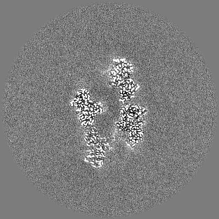
| Images |  emd_70421.png emd_70421.png | 197.8 KB | ||
| Masks | emd_70421_msk_1.map | 347.6 MB | Mask map | |
| Filedesc metadata | emd-70421.cif.gz | 6.9 KB | ||
| Others | emd_70421_additional_1.map.gzemd_70421_half_map_1.map.gzemd_70421_half_map_2.map.gz | 172.3 MB 322.3 MB 322.3 MB | ||
| Archive directory |  http://ftp.pdbj.org/pub/emdb/structures/EMD-70421ftp://ftp.pdbj.org/pub/emdb/structures/EMD-70421 http://ftp.pdbj.org/pub/emdb/structures/EMD-70421ftp://ftp.pdbj.org/pub/emdb/structures/EMD-70421 | HTTPS FTP |
-Related structure data
| Related structure data |  9ofaMC  9odtC C: citing same article ( M: atomic model generated by this map |
|---|---|
| Similar structure data |

-Links
| EMDB pages | EMDB (EBI/PDBe) / EMDataResource |
|---|
-Map
| File | Download / File: emd_70421.map.gz / Format: CCP4 / Size: 347.6 MB / Type: IMAGE STORED AS FLOATING POINT NUMBER (4 BYTES) | ||||||||||||||||||||||||||||||||||||
|---|---|---|---|---|---|---|---|---|---|---|---|---|---|---|---|---|---|---|---|---|---|---|---|---|---|---|---|---|---|---|---|---|---|---|---|---|---|
| Annotation | The structure of a Fungal Cyanide Hydratase from Gloeocercospora sorghi | ||||||||||||||||||||||||||||||||||||
| Projections & slices | Image control
Images are generated by Spider. | ||||||||||||||||||||||||||||||||||||
| Voxel size | X=Y=Z: 0.8748 Å | ||||||||||||||||||||||||||||||||||||
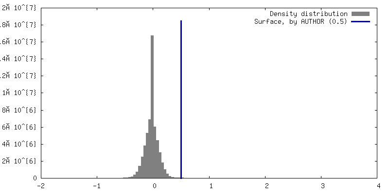
| Density |
| ||||||||||||||||||||||||||||||||||||
| Symmetry | Space group: 1 | ||||||||||||||||||||||||||||||||||||
| Details | EMDB XML:
|
 Z (Sec.)
 Y (Row.)
 X (Col.)


-Supplemental data
-Mask #1
| File | emd_70421_msk_1.map | ||||||||||||
|---|---|---|---|---|---|---|---|---|---|---|---|---|---|
| Projections & Slices |
| ||||||||||||
| Density Histograms |


-Additional map: Map with the helical axis aligend to the...
| File | emd_70421_additional_1.map | ||||||||||||
|---|---|---|---|---|---|---|---|---|---|---|---|---|---|
| Annotation | Map with the helical axis aligend to the Z-axis. This map could have a slightly less resolution compared to the main map. | ||||||||||||
| Projections & Slices |
| ||||||||||||
| Density Histograms |


-Half map: Half map of the structure of a Fungal...
| File | emd_70421_half_map_1.map | ||||||||||||
|---|---|---|---|---|---|---|---|---|---|---|---|---|---|
| Annotation | Half map of the structure of a Fungal Cyanide Hydratase from Gloeocercospora sorghi | ||||||||||||
| Projections & Slices |
| ||||||||||||
| Density Histograms |


-Half map: Half map of the structure of a Fungal...
| File | emd_70421_half_map_2.map | ||||||||||||
|---|---|---|---|---|---|---|---|---|---|---|---|---|---|
| Annotation | Half map of the structure of a Fungal Cyanide Hydratase from Gloeocercospora sorghi | ||||||||||||
| Projections & Slices |
| ||||||||||||
| Density Histograms |


- Sample components
-Entire : Quaternary lef-handed helix of cyanide hydratase dimers
| Entire | Name: Quaternary lef-handed helix of cyanide hydratase dimers |
|---|---|
| Components |
|
-Supramolecule #1: Quaternary lef-handed helix of cyanide hydratase dimers
| Supramolecule | Name: Quaternary lef-handed helix of cyanide hydratase dimers type: complex / ID: 1 / Parent: 0 / Macromolecule list: all / Details: wild type CynH from Gloeocercospora sorghi |
|---|---|
| Source (natural) | Organism: Microdochium sorghi (fungus) |
| Molecular weight | Theoretical: 981.582 KDa |
-Macromolecule #1: Cyanide hydratase
| Macromolecule | Name: Cyanide hydratase / type: protein_or_peptide / ID: 1 / Details: Monomer of CynH / Number of copies: 20 / Enantiomer: LEVO / EC number: cyanide hydratase |
|---|---|
| Source (natural) | Organism: Microdochium sorghi (fungus) |
| Molecular weight | Theoretical: 40.946137 KDa |
| Recombinant expression | Organism:  |
| Sequence | String: MPINKYKAAV VTSEPVWENL EGGVVKTIEF INEAGKAGCK LIAFPEVWIP GYPYWMWKVN YLQSLPMLKA YRENSIAMDS SEMRRIRAA ARDNQIYVSI GVSEIDHATL YLTQVLISPL GDVINHRRKI KPTHVEKLVY GDGSGDSFEP VTQTEIGRLG Q LNCWENMN ...String: MPINKYKAAV VTSEPVWENL EGGVVKTIEF INEAGKAGCK LIAFPEVWIP GYPYWMWKVN YLQSLPMLKA YRENSIAMDS SEMRRIRAA ARDNQIYVSI GVSEIDHATL YLTQVLISPL GDVINHRRKI KPTHVEKLVY GDGSGDSFEP VTQTEIGRLG Q LNCWENMN PFLKSLAVAR GEQIHVAAWP VYPDLSKQVH PDPATNYADP ASDLVTPAYA IETGTWVLAP FQRISVEGLK RH TPPGVEP ETDATPYNGH ARIFRPDGSL YAKPAVDFDG LMYVDIDLNE SHLTKALADF AGHYMRPDLI RLLVDTRRKE LVT EVGGGD NGGIQSYSTM ARLGLDRPLE EEDYRQGTDA GETEKASSNG HA UniProtKB: Cyanide hydratase |
-Experimental details
-Structure determination
| Method | cryo EM |
|---|---|
 Processing Processing | single particle reconstruction |
| Aggregation state | particle |
-Sample preparation
| Concentration | 1.5 mg/mL | |||||||||
|---|---|---|---|---|---|---|---|---|---|---|
| Buffer | pH: 8 Component:
| |||||||||
| Grid | Model: Quantifoil R2/2 / Material: COPPER / Mesh: 200 / Support film - Material: CARBON / Support film - topology: HOLEY / Support film - Film thickness: 12 / Pretreatment - Type: GLOW DISCHARGE / Pretreatment - Time: 25 sec. / Pretreatment - Atmosphere: AIR | |||||||||
| Vitrification | Cryogen name: ETHANE / Chamber humidity: 100 % / Chamber temperature: 277.15 K / Instrument: FEI VITROBOT MARK IV |
- Electron microscopy
| Microscope | TFS KRIOS |
|---|---|
| Image recording | Film or detector model: TFS FALCON 4i (4k x 4k) / Number grids imaged: 1 / Number real images: 5928 / Average exposure time: 4.39 sec. / Average electron dose: 55.59 e/Å2 Details: Number of processing frames fractions collected were 30 |
| Electron beam | Acceleration voltage: 300 kV / Electron source:  FIELD EMISSION GUN FIELD EMISSION GUN |
| Electron optics | Illumination mode: FLOOD BEAM / Imaging mode: BRIGHT FIELD / Nominal defocus max: 2.2 µm / Nominal defocus min: 0.8 µm / Nominal magnification: 165000 |
| Experimental equipment |  Model: Titan Krios / Image courtesy: FEI Company |
+Image processing

-Atomic model buiding 1
| Initial model | Chain - Source name: AlphaFold / Chain - Initial model type: in silico model |
|---|---|
| Output model | PDB-9ofa: |
