 ムービー
ムービー コントローラー


+ データを開く

- 基本情報
基本情報
| 登録情報 | 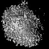 | |||||||||||||||
|---|---|---|---|---|---|---|---|---|---|---|---|---|---|---|---|---|
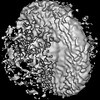
| タイトル | Structure of the human prohibitin complex in the closed state | |||||||||||||||
 マップデータ マップデータ | ||||||||||||||||
 試料 試料 |
| |||||||||||||||
 キーワード キーワード | Mitochondria / Membrane / MEMBRANE PROTEIN | |||||||||||||||
| 機能・相同性 |  機能・相同性情報 機能・相同性情報mitochondrial prohibitin complex / regulation of cardiolipin metabolic process / regulation of cytochrome-c oxidase activity / : / complement component C3a binding / proteinase activated receptor binding / host-mediated perturbation of viral RNA genome replication / regulation of branching involved in mammary gland duct morphogenesis / negative regulation of mammary gland epithelial cell proliferation / Processing of SMDT1 ...mitochondrial prohibitin complex / regulation of cardiolipin metabolic process / regulation of cytochrome-c oxidase activity / : / complement component C3a binding / proteinase activated receptor binding / host-mediated perturbation of viral RNA genome replication / regulation of branching involved in mammary gland duct morphogenesis / negative regulation of mammary gland epithelial cell proliferation / Processing of SMDT1 / sphingolipid binding / negative regulation of nuclear receptor-mediated glucocorticoid signaling pathway / T-helper 17 type immune response / Cellular response to mitochondrial stress / RIG-I signaling pathway / positive regulation of complement activation / mammary gland branching involved in thelarche / complement component C3b binding / negative regulation of intracellular estrogen receptor signaling pathway / : / negative regulation of androgen receptor signaling pathway / positive regulation of G protein-coupled receptor signaling pathway / cellular response to interleukin-6 / mammary gland epithelial cell proliferation / sister chromatid cohesion / positive regulation of immunoglobulin production / DNA biosynthetic process / positive regulation of interleukin-17 production / progesterone receptor signaling pathway / B cell activation / mammary gland alveolus development / : / : / mitophagy / estrogen receptor signaling pathway / epigenetic regulation of gene expression / positive regulation of smooth muscle cell proliferation / antiviral innate immune response / nuclear estrogen receptor binding / cell periphery / mitochondrion organization / RAF activation / negative regulation of cell growth / positive regulation of non-canonical NF-kappaB signal transduction / negative regulation of protein catabolic process / negative regulation of ERK1 and ERK2 cascade / histone deacetylase binding / nuclear matrix / protein import into nucleus / osteoblast differentiation / Signaling by moderate kinase activity BRAF mutants / Paradoxical activation of RAF signaling by kinase inactive BRAF / Signaling downstream of RAS mutants / transcription corepressor activity / positive regulation of neuron apoptotic process / cell migration / regulation of apoptotic process / early endosome / mitochondrial outer membrane / positive regulation of ERK1 and ERK2 cascade / positive regulation of phosphatidylinositol 3-kinase/protein kinase B signal transduction / mitochondrial inner membrane / protein stabilization / protein heterodimerization activity / negative regulation of cell population proliferation / negative regulation of DNA-templated transcription / symbiont entry into host cell / positive regulation of gene expression / regulation of DNA-templated transcription / negative regulation of apoptotic process / positive regulation of DNA-templated transcription / enzyme binding / cell surface / negative regulation of transcription by RNA polymerase II / signal transduction / protein homodimerization activity / protein-containing complex / mitochondrion / extracellular exosome / nucleoplasm / membrane / identical protein binding / nucleus / plasma membrane / cytoplasm 類似検索 - 分子機能 | |||||||||||||||
| 生物種 |  Homo sapiens (ヒト) Homo sapiens (ヒト) | |||||||||||||||
| 手法 | サブトモグラム平均法 / クライオ電子顕微鏡法 / 解像度: 21.0 Å | |||||||||||||||
 データ登録者 データ登録者 | Rose K / Herrmann E / Hurley JH | |||||||||||||||
| 資金援助 |  米国, 米国,  ドイツ, 4件 ドイツ, 4件
| |||||||||||||||
 引用 引用 | ジャーナル: Proc Natl Acad Sci U S A / 年: 2025 タイトル: In situ cryo-ET visualization of mitochondrial depolarization and mitophagic engulfment. 著者: Kevin Rose / Eric Herrmann / Eve Kakudji / Javier Lizarrondo / A Yasemin Celebi / Florian Wilfling / Samantha C Lewis / James H Hurley / 要旨: Defective mitochondrial quality control in response to loss of mitochondrial membrane polarization is implicated in Parkinson's disease by mutations in and . Parkin-expressing U2 osteosarcoma (U2OS) ...Defective mitochondrial quality control in response to loss of mitochondrial membrane polarization is implicated in Parkinson's disease by mutations in and . Parkin-expressing U2 osteosarcoma (U2OS) cells were treated with the depolarizing agents oligomycin and antimycin A (OA) and subjected to cryo-focused ion beam milling and in situ cryo-electron tomography. Mitochondria were fragmented and devoid of matrix calcium phosphate crystals. Phagophores were visualized, with bridge-like lipid transporter densities connected to mitophagic phagophores. A subpopulation of ATP synthases relocalized from cristae to the inner boundary membrane. The structure of the dome-shaped prohibitin complex, a dodecamer of PHB1-PHB2 dimers, was determined in situ by subtomogram averaging in untreated and treated cells and found to exist in open and closed conformations, with the closed conformation being enriched by OA treatment. These findings provide a set of native snapshots of the manifold nano-structural consequences of mitochondrial depolarization and provide a baseline for future in situ dissection of Parkin-dependent mitophagy. | |||||||||||||||
| 履歴 |
|
- 構造の表示
構造の表示
| 添付画像 |
|---|

- ダウンロードとリンク
-EMDBアーカイブ
| マップデータ | emd_70179.map.gz | 6.2 MB | EMDBマップデータ形式 | |
|---|---|---|---|---|
| ヘッダ (付随情報) | emd-70179-v30.xmlemd-70179.xml | 25.6 KB 25.6 KB | 表示 表示 | EMDBヘッダ |
| FSC (解像度算出) | emd_70179_fsc.xml | 4.3 KB | 表示 | FSCデータファイル |
| 画像 |  emd_70179.png emd_70179.png | 48.9 KB | ||
| マスクデータ | emd_70179_msk_1.map | 6.6 MB | マスクマップ | |
| Filedesc metadata | emd-70179.cif.gz | 6.7 KB | ||
| その他 | emd_70179_half_map_1.map.gzemd_70179_half_map_2.map.gz | 5 MB 4.9 MB | ||
| アーカイブディレクトリ |  http://ftp.pdbj.org/pub/emdb/structures/EMD-70179ftp://ftp.pdbj.org/pub/emdb/structures/EMD-70179 http://ftp.pdbj.org/pub/emdb/structures/EMD-70179ftp://ftp.pdbj.org/pub/emdb/structures/EMD-70179 | HTTPS FTP |
-関連構造データ
-リンク
| EMDBのページ | EMDB (EBI/PDBe) / EMDataResource |
|---|---|
| 「今月の分子」の関連する項目 |

-マップ
| ファイル | ダウンロード / ファイル: emd_70179.map.gz / 形式: CCP4 / 大きさ: 6.6 MB / タイプ: IMAGE STORED AS FLOATING POINT NUMBER (4 BYTES) | ||||||||||||||||||||||||||||||||||||
|---|---|---|---|---|---|---|---|---|---|---|---|---|---|---|---|---|---|---|---|---|---|---|---|---|---|---|---|---|---|---|---|---|---|---|---|---|---|
| 投影像・断面図 | 画像のコントロール
画像は Spider により作成 | ||||||||||||||||||||||||||||||||||||
| ボクセルのサイズ | X=Y=Z: 4.2 Å | ||||||||||||||||||||||||||||||||||||
| 密度 |
| ||||||||||||||||||||||||||||||||||||
| 対称性 | 空間群: 1 | ||||||||||||||||||||||||||||||||||||
| 詳細 | EMDB XML:
|
 Z (Sec.)
 Y (Row.)
 X (Col.)


-添付データ
-マスク #1
| ファイル | emd_70179_msk_1.map | ||||||||||||
|---|---|---|---|---|---|---|---|---|---|---|---|---|---|
| 投影像・断面図 |
| ||||||||||||
| 密度ヒストグラム |


-ハーフマップ: #2
| ファイル | emd_70179_half_map_1.map | ||||||||||||
|---|---|---|---|---|---|---|---|---|---|---|---|---|---|
| 投影像・断面図 |
| ||||||||||||
| 密度ヒストグラム |


-ハーフマップ: #1
| ファイル | emd_70179_half_map_2.map | ||||||||||||
|---|---|---|---|---|---|---|---|---|---|---|---|---|---|
| 投影像・断面図 |
| ||||||||||||
| 密度ヒストグラム |


- 試料の構成要素
-全体 : Human prohibitin complex consisting of PHB1 and PHB2
| 全体 | 名称: Human prohibitin complex consisting of PHB1 and PHB2 |
|---|---|
| 要素 |
|
-超分子 #1: Human prohibitin complex consisting of PHB1 and PHB2
| 超分子 | 名称: Human prohibitin complex consisting of PHB1 and PHB2 タイプ: cell / ID: 1 / 親要素: 0 / 含まれる分子: all |
|---|---|
| 由来(天然) | 生物種: Homo sapiens (ヒト) |
-分子 #1: Prohibitin-2
| 分子 | 名称: Prohibitin-2 / タイプ: protein_or_peptide / ID: 1 / コピー数: 12 / 光学異性体: LEVO |
|---|---|
| 由来(天然) | 生物種: Homo sapiens (ヒト) |
| 分子量 | 理論値: 33.341355 KDa |
| 配列 | 文字列: MAQNLKDLAG RLPAGPRGMG TALKLLLGAG AVAYGVRESV FTVEGGHRAI FFNRIGGVQQ DTILAEGLHF RIPWFQYPII YDIRARPRK ISSPTGSKDL QMVNISLRVL SRPNAQELPS MYQRLGLDYE ERVLPSIVNE VLKSVVAKFN ASQLITQRAQ V SLLIRREL ...文字列: MAQNLKDLAG RLPAGPRGMG TALKLLLGAG AVAYGVRESV FTVEGGHRAI FFNRIGGVQQ DTILAEGLHF RIPWFQYPII YDIRARPRK ISSPTGSKDL QMVNISLRVL SRPNAQELPS MYQRLGLDYE ERVLPSIVNE VLKSVVAKFN ASQLITQRAQ V SLLIRREL TERAKDFSLI LDDVAITELS FSREYTAAVE AKQVAQQEAQ RAQFLVEKAK QEQRQKIVQA EGEAEAAKML GE ALSKNPG YIKLRKIRAA QNISKTIATS QNRIYLTADN LVLNLQDESF TRGSDSLIKG KK UniProtKB: Prohibitin-2 |
-分子 #2: Prohibitin 1
| 分子 | 名称: Prohibitin 1 / タイプ: protein_or_peptide / ID: 2 / コピー数: 12 / 光学異性体: LEVO |
|---|---|
| 由来(天然) | 生物種: Homo sapiens (ヒト) |
| 分子量 | 理論値: 29.838029 KDa |
| 配列 | 文字列: MAAKVFESIG KFGLALAVAG GVVNSALYNV DAGHRAVIFD RFRGVQDIVV GEGTHFLIPW VQKPIIFDCR SRPRNVPVIT GSKDLQNVN ITLRILFRPV ASQLPRIFTS IGEDYDERVL PSITTEILKS VVARFDAGEL ITQRELVSRQ VSDDLTERAA T FGLILDDV ...文字列: MAAKVFESIG KFGLALAVAG GVVNSALYNV DAGHRAVIFD RFRGVQDIVV GEGTHFLIPW VQKPIIFDCR SRPRNVPVIT GSKDLQNVN ITLRILFRPV ASQLPRIFTS IGEDYDERVL PSITTEILKS VVARFDAGEL ITQRELVSRQ VSDDLTERAA T FGLILDDV SLTHLTFGKE FTEAVEAKQV AQQEAERARF VVEKAEQQKK AAIISAEGDS KAAELIANSL ATAGDGLIEL RK LEAAEDI AYQLSRSRNI TYLPAGQSVL LQLPQ UniProtKB: Prohibitin 1 |
-実験情報
-構造解析
| 手法 | クライオ電子顕微鏡法 |
|---|---|
 解析 解析 | サブトモグラム平均法 |
| 試料の集合状態 | cell |
-試料調製
| 緩衝液 | pH: 7.4 |
|---|---|
| グリッド | モデル: Quantifoil R2/2 / 材質: GOLD / メッシュ: 200 / 支持フィルム - 材質: CARBON / 支持フィルム - トポロジー: HOLEY / 前処理 - タイプ: GLOW DISCHARGE / 前処理 - 雰囲気: AIR |
| 凍結 | 凍結剤: ETHANE / チャンバー内湿度: 90 % / チャンバー内温度: 310.15 K / 装置: FEI VITROBOT MARK IV |
- 電子顕微鏡法 #1
| Microscopy ID | 1 |
|---|---|
| 顕微鏡 | TFS KRIOS |
| 特殊光学系 | エネルギーフィルター - 名称: GIF Bioquantum / エネルギーフィルター - スリット幅: 25 eV |
| 撮影 | Image recording ID: 1 フィルム・検出器のモデル: GATAN K3 BIOQUANTUM (6k x 4k) 平均電子線量: 2.1 e/Å2 |
| 電子線 | 加速電圧: 300 kV / 電子線源:  FIELD EMISSION GUN FIELD EMISSION GUN |
| 電子光学系 | 照射モード: FLOOD BEAM / 撮影モード: BRIGHT FIELD / 最大 デフォーカス(公称値): 6.0 µm / 最小 デフォーカス(公称値): 2.0 µm / 倍率(公称値): 43000 |
| 試料ステージ | 試料ホルダーモデル: FEI TITAN KRIOS AUTOGRID HOLDER ホルダー冷却材: NITROGEN |
| 実験機器 |  モデル: Titan Krios / 画像提供: FEI Company |
-電子顕微鏡法 #1~
| Microscopy ID | 1 |
|---|---|
| 顕微鏡 | TFS KRIOS |
| 特殊光学系 | エネルギーフィルター - 名称: TFS Selectris X / エネルギーフィルター - スリット幅: 10 eV |
| 撮影 | Image recording ID: 2 フィルム・検出器のモデル: FEI FALCON IV (4k x 4k) 平均電子線量: 3.0 e/Å2 |
| 電子線 | 加速電圧: 300 kV / 電子線源: FIELD EMISSION GUN |
| 電子光学系 | 照射モード: FLOOD BEAM / 撮影モード: BRIGHT FIELD / 最大 デフォーカス(公称値): 6.0 µm / 最小 デフォーカス(公称値): 2.0 µm / 倍率(公称値): 64000 |
| 試料ステージ | 試料ホルダーモデル: FEI TITAN KRIOS AUTOGRID HOLDER ホルダー冷却材: NITROGEN |
| 実験機器 | モデル: Titan Krios / 画像提供: FEI Company |
-電子顕微鏡法 #1~~
| Microscopy ID | 1 |
|---|---|
| 顕微鏡 | TFS KRIOS |
| 特殊光学系 | エネルギーフィルター - 名称: GIF Bioquantum / エネルギーフィルター - スリット幅: 20 eV |
| 撮影 | Image recording ID: 3 フィルム・検出器のモデル: GATAN K3 BIOQUANTUM (6k x 4k) 平均電子線量: 3.0 e/Å2 |
| 電子線 | 加速電圧: 300 kV / 電子線源: FIELD EMISSION GUN |
| 電子光学系 | 照射モード: FLOOD BEAM / 撮影モード: BRIGHT FIELD / 最大 デフォーカス(公称値): 6.0 µm / 最小 デフォーカス(公称値): 2.0 µm / 倍率(公称値): 42000 |
| 試料ステージ | 試料ホルダーモデル: FEI TITAN KRIOS AUTOGRID HOLDER ホルダー冷却材: NITROGEN |
| 実験機器 | モデル: Titan Krios / 画像提供: FEI Company |
+画像解析

-原子モデル構築 1
| 初期モデル | Chain - Source name: AlphaFold / Chain - Initial model type: in silico model |
|---|---|
| 精密化 | 空間: REAL / プロトコル: AB INITIO MODEL |
| 得られたモデル |  PDB-9o6s: |
