Movie
Movie Controller

[English] 日本語
 Yorodumi
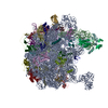
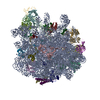
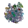
- EMDB-7005: Negative stain reconstruction of the peroxisomal AAA-ATPase Pex1/... -
+ Open data

- Basic information
| Entry | Database: EMDB / ID: EMD-7005 | ||||||||||||
|---|---|---|---|---|---|---|---|---|---|---|---|---|---|
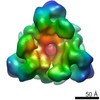
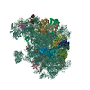
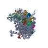
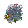
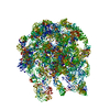
| Title | Negative stain reconstruction of the peroxisomal AAA-ATPase Pex1/Pex6 complex associated with substrate Pex15 | ||||||||||||
 Map data Map data | Negative stain reconstruction of the Pex15 bound Pex1-Pex6 complex. | ||||||||||||
 Sample Sample |
| ||||||||||||
| Biological species |  | ||||||||||||
| Method | single particle reconstruction / negative staining / Resolution: 23.2 Å | ||||||||||||
 Authors Authors | Chowdhury S / Gardner BM / Castanzo DT / Stjepanovic G / Stefely MS / Hurley JH / Martin A / Lander GC | ||||||||||||
| Funding support |  United States, 3 items United States, 3 items
| ||||||||||||
 Citation Citation | Journal: Nat Commun / Year: 2018 Title: The peroxisomal AAA-ATPase Pex1/Pex6 unfolds substrates by processive threading. Authors: Brooke M Gardner / Dominic T Castanzo / Saikat Chowdhury / Goran Stjepanovic / Matthew S Stefely / James H Hurley / Gabriel C Lander / Andreas Martin / Abstract: Pex1 and Pex6 form a heterohexameric motor essential for peroxisome biogenesis and function, and mutations in these AAA-ATPases cause most peroxisome-biogenesis disorders in humans. The tail-anchored ...Pex1 and Pex6 form a heterohexameric motor essential for peroxisome biogenesis and function, and mutations in these AAA-ATPases cause most peroxisome-biogenesis disorders in humans. The tail-anchored protein Pex15 recruits Pex1/Pex6 to the peroxisomal membrane, where it performs an unknown function required for matrix-protein import. Here we determine that Pex1/Pex6 from S. cerevisiae is a protein translocase that unfolds Pex15 in a pore-loop-dependent and ATP-hydrolysis-dependent manner. Our structural studies of Pex15 in isolation and in complex with Pex1/Pex6 illustrate that Pex15 binds the N-terminal domains of Pex6, before its C-terminal disordered region engages with the pore loops of the motor, which then processively threads Pex15 through the central pore. Furthermore, Pex15 directly binds the cargo receptor Pex5, linking Pex1/Pex6 to other components of the peroxisomal import machinery. Our results thus support a role of Pex1/Pex6 in mechanical unfolding of peroxins or their extraction from the peroxisomal membrane during matrix-protein import. | ||||||||||||
| History |
|
- Structure visualization
| Movie |
 Movie viewer Movie viewer |
|---|---|
| Structure viewer | EM map: SurfViewMolmilJmol/JSmol |
| Supplemental images |
 UCSF Chimera
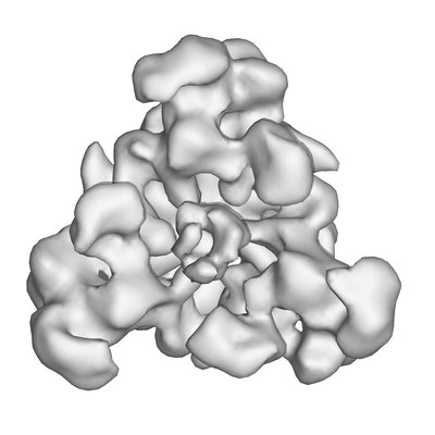
- Downloads & links
-EMDB archive
| Map data | emd_7005.map.gz | 1.8 MB | EMDB map data format | |
|---|---|---|---|---|
| Header (meta data) | emd-7005-v30.xmlemd-7005.xml | 12.5 KB 12.5 KB | Display Display | EMDB header |
| Images |  emd_7005.png emd_7005.png | 58.5 KB | ||
| Archive directory |  http://ftp.pdbj.org/pub/emdb/structures/EMD-7005ftp://ftp.pdbj.org/pub/emdb/structures/EMD-7005 http://ftp.pdbj.org/pub/emdb/structures/EMD-7005ftp://ftp.pdbj.org/pub/emdb/structures/EMD-7005 | HTTPS FTP |
-Validation report
| Summary document | emd_7005_validation.pdf.gz | 78.8 KB | Display | EMDB validaton report |
|---|---|---|---|---|
| Full document | emd_7005_full_validation.pdf.gz | 77.9 KB | Display | |
| Data in XML | emd_7005_validation.xml.gz | 493 B | Display | |
| Arichive directory | https://ftp.pdbj.org/pub/emdb/validation_reports/EMD-7005ftp://ftp.pdbj.org/pub/emdb/validation_reports/EMD-7005 | HTTPS FTP |
-Related structure data
-Links
| EMDB pages | EMDB (EBI/PDBe) / EMDataResource |
|---|
-Map
| File | Download / File: emd_7005.map.gz / Format: CCP4 / Size: 2 MB / Type: IMAGE STORED AS FLOATING POINT NUMBER (4 BYTES) | ||||||||||||||||||||||||||||||||||||||||||||||||||||||||||||||||||||
|---|---|---|---|---|---|---|---|---|---|---|---|---|---|---|---|---|---|---|---|---|---|---|---|---|---|---|---|---|---|---|---|---|---|---|---|---|---|---|---|---|---|---|---|---|---|---|---|---|---|---|---|---|---|---|---|---|---|---|---|---|---|---|---|---|---|---|---|---|---|
| Annotation | Negative stain reconstruction of the Pex15 bound Pex1-Pex6 complex. | ||||||||||||||||||||||||||||||||||||||||||||||||||||||||||||||||||||
| Projections & slices | Image control
Images are generated by Spider. | ||||||||||||||||||||||||||||||||||||||||||||||||||||||||||||||||||||
| Voxel size | X=Y=Z: 4.1 Å | ||||||||||||||||||||||||||||||||||||||||||||||||||||||||||||||||||||
| Density |
| ||||||||||||||||||||||||||||||||||||||||||||||||||||||||||||||||||||
| Symmetry | Space group: 1 | ||||||||||||||||||||||||||||||||||||||||||||||||||||||||||||||||||||
| Details | EMDB XML:
CCP4 map header:
| ||||||||||||||||||||||||||||||||||||||||||||||||||||||||||||||||||||
 Z (Sec.)
 Y (Row.)
 X (Col.)


-Supplemental data
- Sample components
-Entire : Complex between Pex1-Pex6 AAA-ATPase with substrate Pex15
| Entire | Name: Complex between Pex1-Pex6 AAA-ATPase with substrate Pex15 |
|---|---|
| Components |
|
-Supramolecule #1: Complex between Pex1-Pex6 AAA-ATPase with substrate Pex15
| Supramolecule | Name: Complex between Pex1-Pex6 AAA-ATPase with substrate Pex15 type: complex / ID: 1 / Parent: 0 |
|---|---|
| Source (natural) | Organism: |
| Recombinant expression | Organism:  |
| Molecular weight | Theoretical: 750 KDa |
-Experimental details
-Structure determination
| Method | negative staining |
|---|---|
 Processing Processing | single particle reconstruction |
| Aggregation state | particle |
-Sample preparation
| Buffer | pH: 7.6 Details: 60 mM HEPES pH 7.6, 50 mM NaCl, 50 mM KCl, 10 % glycerol, 10 mM MgCl2, 0.5 mM EDTA and 5mM ATP |
|---|---|
| Staining | Type: NEGATIVE / Material: Uranyl Formate |
| Grid | Model: Maxtaform / Material: COPPER/RHODIUM / Mesh: 400 / Support film - Material: CARBON / Pretreatment - Type: GLOW DISCHARGE / Pretreatment - Atmosphere: AIR |
- Electron microscopy
| Microscope | FEI TECNAI SPIRIT |
|---|---|
| Temperature | Max: 298.15 K |
| Image recording | Film or detector model: TVIPS TEMCAM-F416 (4k x 4k) / Digitization - Dimensions - Width: 4096 pixel / Digitization - Dimensions - Height: 4096 pixel / Digitization - Sampling interval: 15.6 µm / Number grids imaged: 1 / Number real images: 1097 / Average exposure time: 0.4 sec. / Average electron dose: 20.0 e/Å2 |
| Electron beam | Acceleration voltage: 120 kV / Electron source: LAB6 |
| Electron optics | Illumination mode: FLOOD BEAM / Imaging mode: BRIGHT FIELD / Cs: 2.2 mm / Nominal defocus max: 1.5 µm / Nominal defocus min: 0.5 µm / Nominal magnification: 52000 |
| Sample stage | Specimen holder model: SIDE ENTRY, EUCENTRIC |
| Experimental equipment |  Model: Tecnai Spirit / Image courtesy: FEI Company |