[English] 日本語
Yorodumi
- EMDB-7005: Negative stain reconstruction of the peroxisomal AAA-ATPase Pex1/... -

+
Open data


ID or keywords:

Loading...

-
Basic information

Entry
Database: EMDB / ID: EMD-7005
TitleNegative stain reconstruction of the peroxisomal AAA-ATPase Pex1/Pex6 complex associated with substrate Pex15
Map dataNegative stain reconstruction of the Pex15 bound Pex1-Pex6 complex.
Sample
  • Complex: Complex between Pex1-Pex6 AAA-ATPase with substrate Pex15
Biological speciesSaccharomyces cerevisiae (brewer's yeast)
Methodsingle particle reconstruction / negative staining / Resolution: 23.2 Å
AuthorsChowdhury S / Gardner BM / Castanzo DT / Stjepanovic G / Stefely MS / Hurley JH / Martin A / Lander GC
Funding support United States, 3 items
OrganizationGrant numberCountry
National Institutes of Health/National Institute of General Medical Sciences (NIH/NIGMS)R01-GM094497, NSF-MCB- 1150288, DP2 EB020402-01, K99GM121880 United States
Howard Hughes Medical Institute (HHMI) United States
Department of Energy (DOE, United States) United States
Citation
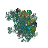
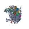
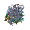
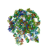
Journal: Nat Commun / Year: 2018
Title: The peroxisomal AAA-ATPase Pex1/Pex6 unfolds substrates by processive threading.
Authors: Brooke M Gardner / Dominic T Castanzo / Saikat Chowdhury / Goran Stjepanovic / Matthew S Stefely / James H Hurley / Gabriel C Lander / Andreas Martin /
Abstract: Pex1 and Pex6 form a heterohexameric motor essential for peroxisome biogenesis and function, and mutations in these AAA-ATPases cause most peroxisome-biogenesis disorders in humans. The tail-anchored ...Pex1 and Pex6 form a heterohexameric motor essential for peroxisome biogenesis and function, and mutations in these AAA-ATPases cause most peroxisome-biogenesis disorders in humans. The tail-anchored protein Pex15 recruits Pex1/Pex6 to the peroxisomal membrane, where it performs an unknown function required for matrix-protein import. Here we determine that Pex1/Pex6 from S. cerevisiae is a protein translocase that unfolds Pex15 in a pore-loop-dependent and ATP-hydrolysis-dependent manner. Our structural studies of Pex15 in isolation and in complex with Pex1/Pex6 illustrate that Pex15 binds the N-terminal domains of Pex6, before its C-terminal disordered region engages with the pore loops of the motor, which then processively threads Pex15 through the central pore. Furthermore, Pex15 directly binds the cargo receptor Pex5, linking Pex1/Pex6 to other components of the peroxisomal import machinery. Our results thus support a role of Pex1/Pex6 in mechanical unfolding of peroxins or their extraction from the peroxisomal membrane during matrix-protein import.
History
DepositionSep 1, 2017-
Header (metadata) releaseDec 20, 2017-
Map releaseDec 20, 2017-
UpdateDec 11, 2019-
Current statusDec 11, 2019Processing site: RCSB / Status: Released

-
Structure visualization

Movie
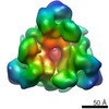
  • Surface view with section colored by density value
  • Surface level: 3.06
  • Imaged by UCSF Chimera
  • Download
  • Surface view colored by cylindrical radius
  • Surface level: 3.06
  • Imaged by UCSF Chimera
  • Download
Movie viewer
Structure viewerEM map:
SurfViewMolmilJmol/JSmol
Supplemental images

Downloads & links

-
Map

FileDownload / File: emd_7005.map.gz / Format: CCP4 / Size: 2 MB / Type: IMAGE STORED AS FLOATING POINT NUMBER (4 BYTES)
AnnotationNegative stain reconstruction of the Pex15 bound Pex1-Pex6 complex.
Voxel sizeX=Y=Z: 4.1 Å
Density
Contour LevelBy AUTHOR: 3.06 / Movie #1: 3.06
Minimum - Maximum-8.168757 - 10.292679
Average (Standard dev.)-0.048110332 (±1.2685611)
SymmetrySpace group: 1
Details

EMDB XML:

Map geometry
Axis orderXYZ
Origin000
Dimensions808080
Spacing808080
CellA=B=C: 328.0 Å
α=β=γ: 90.0 °

CCP4 map header:

modeImage stored as Reals
Å/pix. X/Y/Z4.14.14.1
M x/y/z808080
origin x/y/z0.0000.0000.000
length x/y/z328.000328.000328.000
α/β/γ90.00090.00090.000
start NX/NY/NZ-51-35-11
NX/NY/NZ11110799
MAP C/R/S123
start NC/NR/NS000
NC/NR/NS808080
D min/max/mean-8.16910.293-0.048

-
Supplemental data

-
Sample components

-
Entire : Complex between Pex1-Pex6 AAA-ATPase with substrate Pex15

EntireName: Complex between Pex1-Pex6 AAA-ATPase with substrate Pex15
Components
  • Complex: Complex between Pex1-Pex6 AAA-ATPase with substrate Pex15

-
Supramolecule #1: Complex between Pex1-Pex6 AAA-ATPase with substrate Pex15

SupramoleculeName: Complex between Pex1-Pex6 AAA-ATPase with substrate Pex15
type: complex / ID: 1 / Parent: 0
Source (natural)Organism: Saccharomyces cerevisiae (brewer's yeast)
Recombinant expressionOrganism: Escherichia coli (E. coli) / Recombinant strain: BL21
Molecular weightTheoretical: 750 KDa

-
Experimental details

-
Structure determination

Methodnegative staining
Processingsingle particle reconstruction
Aggregation stateparticle

-
Sample preparation

BufferpH: 7.6
Details: 60 mM HEPES pH 7.6, 50 mM NaCl, 50 mM KCl, 10 % glycerol, 10 mM MgCl2, 0.5 mM EDTA and 5mM ATP
StainingType: NEGATIVE / Material: Uranyl Formate
GridModel: Maxtaform / Material: COPPER/RHODIUM / Mesh: 400 / Support film - Material: CARBON / Pretreatment - Type: GLOW DISCHARGE / Pretreatment - Atmosphere: AIR

-
Electron microscopy

MicroscopeFEI TECNAI SPIRIT
Electron beamAcceleration voltage: 120 kV / Electron source: LAB6
Electron opticsIllumination mode: FLOOD BEAM / Imaging mode: BRIGHT FIELDBright-field microscopy / Cs: 2.2 mm / Nominal defocus max: 1.5 µm / Nominal defocus min: 0.5 µm / Nominal magnification: 52000
Sample stageSpecimen holder model: SIDE ENTRY, EUCENTRIC
TemperatureMax: 298.15 K
Image recordingFilm or detector model: TVIPS TEMCAM-F416 (4k x 4k) / Digitization - Dimensions - Width: 4096 pixel / Digitization - Dimensions - Height: 4096 pixel / Digitization - Sampling interval: 15.6 µm / Number grids imaged: 1 / Number real images: 1097 / Average exposure time: 0.4 sec. / Average electron dose: 20.0 e/Å2
Experimental equipment
Model: Tecnai Spirit / Image courtesy: FEI Company

-
Image processing

Particle selectionNumber selected: 102101
CTF correctionSoftware - Name: CTFFIND3 / Software - details: implemented in Appion
Details: EMAN1 was used for phase flipping whole micrograph before particle extraction.
Startup modelType of model: EMDB MAP
EMDB ID:

Details: This map was low passed filtered to 60 Angstrom resolution.
Initial angle assignmentType: PROJECTION MATCHING
Projection matching processing - Number reference projections: 78118
Software - Name: RELION (ver. 1.3)
Final angle assignmentType: PROJECTION MATCHING / Software - Name: RELION (ver. 1.3)
Final reconstructionApplied symmetry - Point group: C1 (asymmetric) / Resolution.type: BY AUTHOR / Resolution: 23.2 Å / Resolution method: FSC 0.143 CUT-OFF / Software - Name: RELION (ver. 1.3) / Number images used: 14678
DetailsDifference of Gaussians (DoG)-based automated particle picker, implemented in Appion image processing software was used

+
About Yorodumi

-
News

-
Feb 9, 2022. New format data for meta-information of EMDB entries

New format data for meta-information of EMDB entries

  • Version 3 of the EMDB header file is now the official format.
  • The previous official version 1.9 will be removed from the archive.

Related info.:EMDB header

External links:wwPDB to switch to version 3 of the EMDB data model

-
Aug 12, 2020. Covid-19 info

Covid-19 info

URL: https://pdbj.org/emnavi/covid19.php

New page: Covid-19 featured information page in EM Navigator.

Related info.:Covid-19 info / Mar 5, 2020. Novel coronavirus structure data

+
Mar 5, 2020. Novel coronavirus structure data

Novel coronavirus structure data

Related info.:Yorodumi Speices / Aug 12, 2020. Covid-19 info

External links:COVID-19 featured content - PDBj / Molecule of the Month (242):Coronavirus Proteases

+
Jan 31, 2019. EMDB accession codes are about to change! (news from PDBe EMDB page)

EMDB accession codes are about to change! (news from PDBe EMDB page)

  • The allocation of 4 digits for EMDB accession codes will soon come to an end. Whilst these codes will remain in use, new EMDB accession codes will include an additional digit and will expand incrementally as the available range of codes is exhausted. The current 4-digit format prefixed with “EMD-” (i.e. EMD-XXXX) will advance to a 5-digit format (i.e. EMD-XXXXX), and so on. It is currently estimated that the 4-digit codes will be depleted around Spring 2019, at which point the 5-digit format will come into force.
  • The EM Navigator/Yorodumi systems omit the EMD- prefix.

Related info.:Q: What is EMD? / ID/Accession-code notation in Yorodumi/EM Navigator

External links:EMDB Accession Codes are Changing Soon! / Contact to PDBj

+
Jul 12, 2017. Major update of PDB

Major update of PDB

  • wwPDB released updated PDB data conforming to the new PDBx/mmCIF dictionary.
  • This is a major update changing the version number from 4 to 5, and with Remediation, in which all the entries are updated.
  • In this update, many items about electron microscopy experimental information are reorganized (e.g. em_software).
  • Now, EM Navigator and Yorodumi are based on the updated data.

External links:wwPDB Remediation / Enriched Model Files Conforming to OneDep Data Standards Now Available in the PDB FTP Archive

-
Yorodumi

Thousand views of thousand structures

  • Yorodumi is a browser for structure data from EMDB, PDB, SASBDB, etc.
  • This page is also the successor to EM Navigator detail page, and also detail information page/front-end page for Omokage search.
  • The word "yorodu" (or yorozu) is an old Japanese word meaning "ten thousand". "mi" (miru) is to see.

Related info.:EMDB / PDB / SASBDB / Comparison of 3 databanks / Yorodumi Search / Aug 31, 2016. New EM Navigator & Yorodumi / Yorodumi Papers / Jmol/JSmol / Function and homology information / Changes in new EM Navigator and Yorodumi

Read more