 ムービー
ムービー コントローラー


+ データを開く

- 基本情報
基本情報
| 登録情報 | データベース: EMDB / ID: EMD-6948 | |||||||||||||||
|---|---|---|---|---|---|---|---|---|---|---|---|---|---|---|---|---|
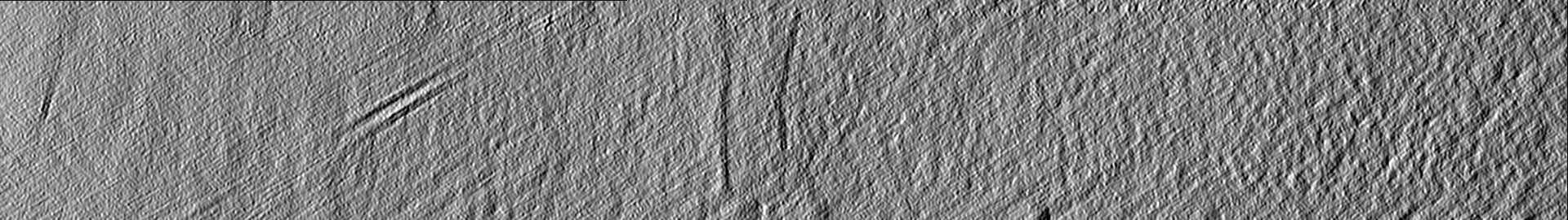
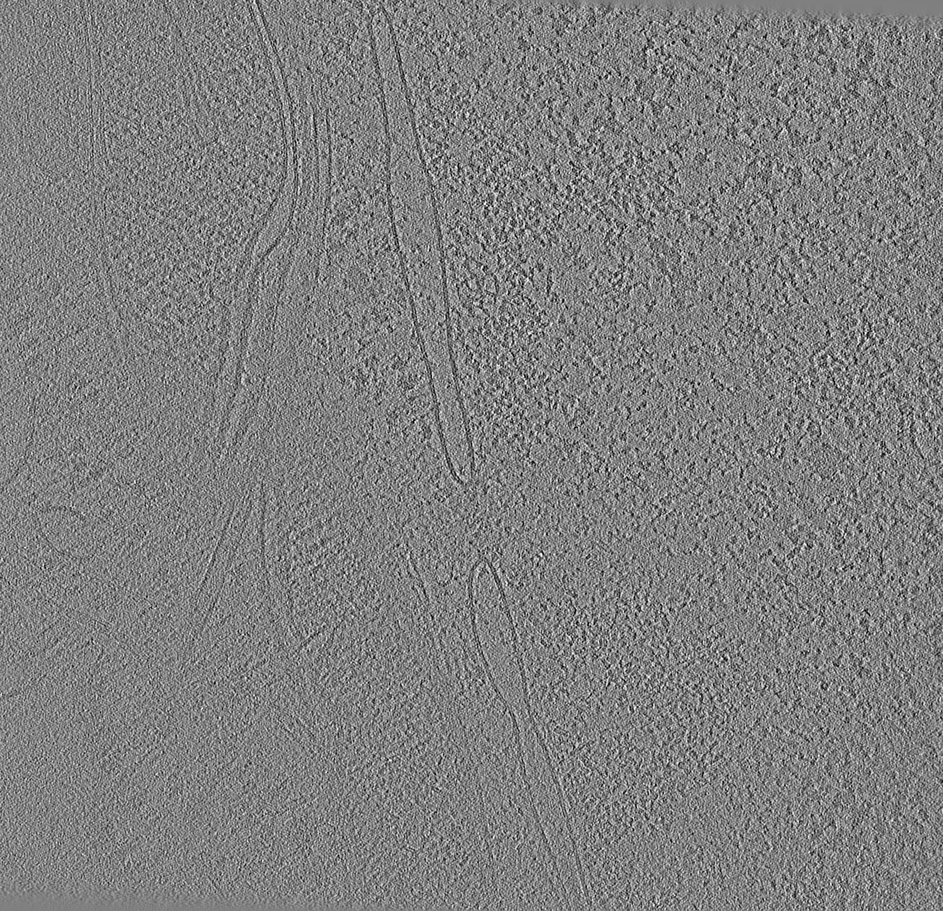
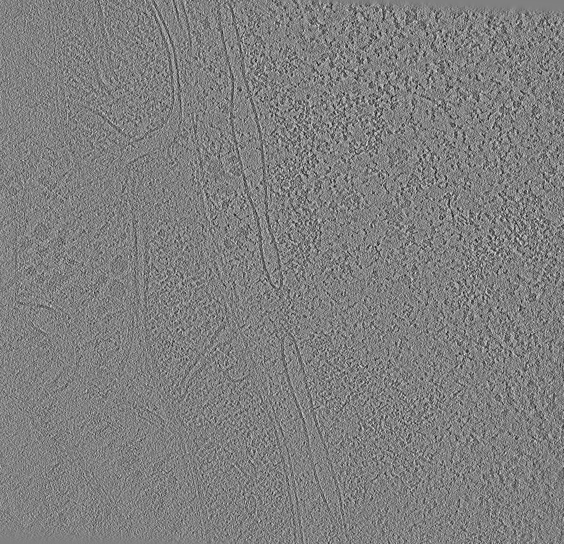
| タイトル | Volta phase contrast cryotomogram of a cryo-FIB lamella of an interphase HeLa cell | |||||||||||||||
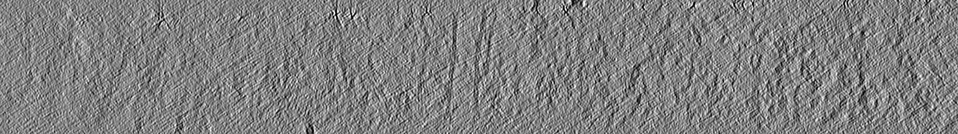
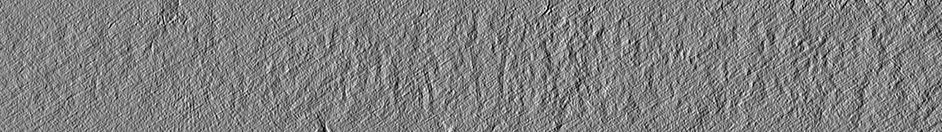
 マップデータ マップデータ | Tomogram of a HeLa cell cryo-FIB lamella. | |||||||||||||||
 試料 試料 |
| |||||||||||||||
| 生物種 |  Homo sapiens (ヒト) Homo sapiens (ヒト) | |||||||||||||||
| 手法 | 電子線トモグラフィー法 / クライオ電子顕微鏡法 | |||||||||||||||
 データ登録者 データ登録者 | Cai S / Bock D / Pilhofer M / Gan L | |||||||||||||||
| 資金援助 |  シンガポール, シンガポール,  スイス, 4件 スイス, 4件
| |||||||||||||||
 引用 引用 | ジャーナル: Mol Biol Cell / 年: 2018 タイトル: The in situ structures of mono-, di-, and trinucleosomes in human heterochromatin. 著者: Shujun Cai / Désirée Böck / Martin Pilhofer / Lu Gan / 要旨: The in situ three-dimensional organization of chromatin at the nucleosome and oligonucleosome levels is unknown. Here we use cryo-electron tomography to determine the in situ structures of HeLa ...The in situ three-dimensional organization of chromatin at the nucleosome and oligonucleosome levels is unknown. Here we use cryo-electron tomography to determine the in situ structures of HeLa nucleosomes, which have canonical core structures and asymmetric, flexible linker DNA. Subtomogram remapping suggests that sequential nucleosomes in heterochromatin follow irregular paths at the oligonucleosome level. This basic principle of higher-order repressive chromatin folding is compatible with the conformational variability of the two linker DNAs at the single-nucleosome level. | |||||||||||||||
| 履歴 |
|
- 構造の表示
構造の表示
| ムービー |
 ムービービューア ムービービューア |
|---|---|
| 添付画像 |

- ダウンロードとリンク
-EMDBアーカイブ
| マップデータ | emd_6948.map.gz | 1.4 GB | EMDBマップデータ形式 | |
|---|---|---|---|---|
| ヘッダ (付随情報) | emd-6948-v30.xmlemd-6948.xml | 11.2 KB 11.2 KB | 表示 表示 | EMDBヘッダ |
| 画像 |  emd_6948.png emd_6948.png | 119.5 KB | ||
| アーカイブディレクトリ |  http://ftp.pdbj.org/pub/emdb/structures/EMD-6948ftp://ftp.pdbj.org/pub/emdb/structures/EMD-6948 http://ftp.pdbj.org/pub/emdb/structures/EMD-6948ftp://ftp.pdbj.org/pub/emdb/structures/EMD-6948 | HTTPS FTP |
-関連構造データ
| 関連構造データ |  6949C  6950C C: 同じ文献を引用 ( |
|---|---|
| 電子顕微鏡画像生データ | EMPIAR-10179 (タイトル: The in situ structures of mono-, di-, and trinucleosomes in human heterochromatin Data size: 1.9 Data #1: Tilt series of a cryo-FIB lamella of an interphase HeLa cell [tilt series]) |
-リンク
| EMDBのページ | EMDB (EBI/PDBe) / EMDataResource |
|---|
-マップ
| ファイル | ダウンロード / ファイル: emd_6948.map.gz / 形式: CCP4 / 大きさ: 1.8 GB / タイプ: IMAGE STORED AS SIGNED INTEGER (2 BYTES) | ||||||||||||||||||||||||||||||||||||||||||||||||||||||||||||||||||||
|---|---|---|---|---|---|---|---|---|---|---|---|---|---|---|---|---|---|---|---|---|---|---|---|---|---|---|---|---|---|---|---|---|---|---|---|---|---|---|---|---|---|---|---|---|---|---|---|---|---|---|---|---|---|---|---|---|---|---|---|---|---|---|---|---|---|---|---|---|---|
| 注釈 | Tomogram of a HeLa cell cryo-FIB lamella. | ||||||||||||||||||||||||||||||||||||||||||||||||||||||||||||||||||||
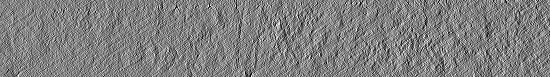
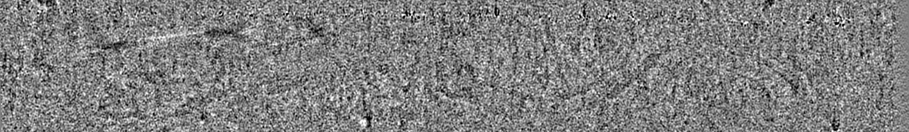
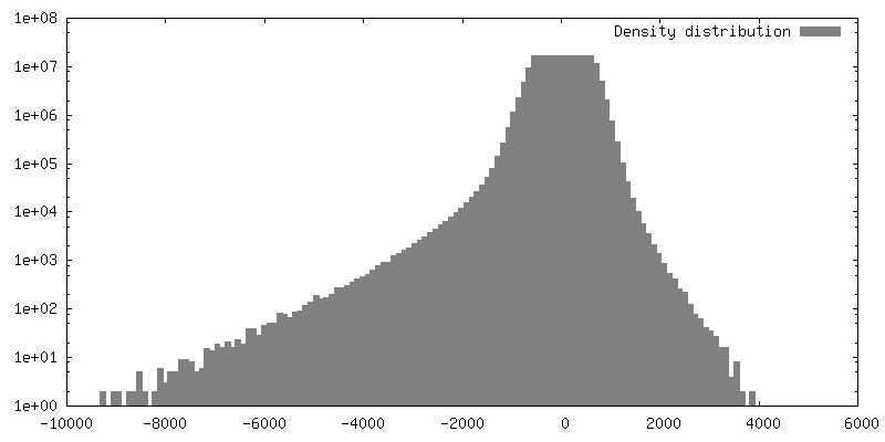
| 投影像・断面図 | 画像のコントロール
画像は Spider により作成 これらの図は立方格子座標系で作成されたものです | ||||||||||||||||||||||||||||||||||||||||||||||||||||||||||||||||||||
| ボクセルのサイズ | X=Y=Z: 6.9 Å | ||||||||||||||||||||||||||||||||||||||||||||||||||||||||||||||||||||
| 密度 |
| ||||||||||||||||||||||||||||||||||||||||||||||||||||||||||||||||||||
| 対称性 | 空間群: 1 | ||||||||||||||||||||||||||||||||||||||||||||||||||||||||||||||||||||
| 詳細 | EMDB XML:
CCP4マップ ヘッダ情報:
| ||||||||||||||||||||||||||||||||||||||||||||||||||||||||||||||||||||
 Z (Sec.)
 Y (Row.)
 X (Col.)


-添付データ
- 試料の構成要素
-全体 : HeLa cell, interphase
| 全体 | 名称: HeLa cell, interphase |
|---|---|
| 要素 |
|
-超分子 #1: HeLa cell, interphase
| 超分子 | 名称: HeLa cell, interphase / タイプ: cell / ID: 1 / 親要素: 0 |
|---|---|
| 由来(天然) | 生物種: Homo sapiens (ヒト) / 器官: cervix |
-実験情報
-構造解析
| 手法 | クライオ電子顕微鏡法 |
|---|---|
 解析 解析 | 電子線トモグラフィー法 |
| 試料の集合状態 | cell |
-試料調製
| 緩衝液 | pH: 7 / 詳細: yeast-extract supplemented |
|---|---|
| グリッド | モデル: Quantifoil R2/2 / 材質: GOLD / メッシュ: 200 / 支持フィルム - 材質: CARBON / 支持フィルム - トポロジー: HOLEY |
| 凍結 | 凍結剤: ETHANE-PROPANE |
| 切片作成 | 集束イオンビーム - 装置: OTHER / 集束イオンビーム - イオン: OTHER / 集束イオンビーム - 電圧: 30 kV / 集束イオンビーム - 電流: 0.025 nA / 集束イオンビーム - Dose rate: 1 ions/(cm2*s) / 集束イオンビーム - 時間: 1 sec. / 集束イオンビーム - 温度: 119 K / 集束イオンビーム - Initial thickness: 2000 nm / 集束イオンビーム - 最終 厚さ: 200 nm 集束イオンビーム - 詳細: The value given for _emd_sectioning_focused_ion_beam.instrument is Helios NanoLab600i. This is not in a list of allowed values set(['DB235', 'OTHER']) so OTHER is ...集束イオンビーム - 詳細: The value given for _emd_sectioning_focused_ion_beam.instrument is Helios NanoLab600i. This is not in a list of allowed values set(['DB235', 'OTHER']) so OTHER is written into the XML file. |
- 電子顕微鏡法
| 顕微鏡 | FEI TITAN KRIOS |
|---|---|
| 特殊光学系 | 位相板: VOLTA PHASE PLATE / エネルギーフィルター - 名称: GIF Quantum LS エネルギーフィルター - エネルギー下限: 0 eV エネルギーフィルター - エネルギー上限: 20 eV |
| 撮影 | フィルム・検出器のモデル: GATAN K2 SUMMIT (4k x 4k) 検出モード: COUNTING / デジタル化 - サイズ - 横: 3838 pixel / デジタル化 - サイズ - 縦: 3710 pixel / デジタル化 - サンプリング間隔: 5.0 µm / 撮影したグリッド数: 1 / 実像数: 61 / 平均電子線量: 2.0 e/Å2 |
| 電子線 | 加速電圧: 300 kV / 電子線源:  FIELD EMISSION GUN FIELD EMISSION GUN |
| 電子光学系 | 倍率(補正後): 14493 / 照射モード: FLOOD BEAM / 撮影モード: BRIGHT FIELD / 最小 デフォーカス(公称値): 0.0 µm |
| 試料ステージ | 試料ホルダーモデル: FEI TITAN KRIOS AUTOGRID HOLDER ホルダー冷却材: NITROGEN |
| 実験機器 |  モデル: Titan Krios / 画像提供: FEI Company |
-画像解析
| 最終 再構成 | アルゴリズム: BACK PROJECTION / ソフトウェア - 名称: IMOD (ver. 4.9) / 使用した粒子像数: 61 |
|---|
