 Movie
Movie Controller

[English] 日本語
 Yorodumi
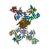
- EMDB-6841: Complex structure of Pseudorabies virus glycoprotein B bound by a... -
+ Open data

- Basic information
| Entry | Database: EMDB / ID: EMD-6841 | |||||||||
|---|---|---|---|---|---|---|---|---|---|---|
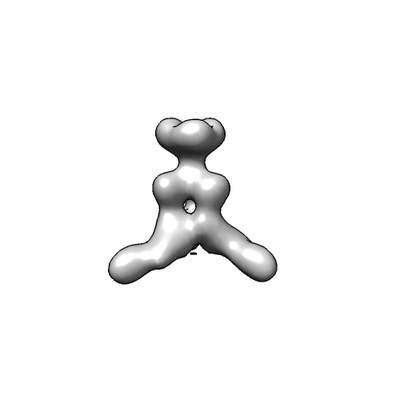
| Title | Complex structure of Pseudorabies virus glycoprotein B bound by a neutralizing antibody | |||||||||
 Map data Map data | EM density map | |||||||||
 Sample Sample |
| |||||||||
| Biological species |  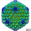 Suid herpesvirus 1 Suid herpesvirus 1 | |||||||||
| Method | single particle reconstruction / negative staining / Resolution: 35.0 Å | |||||||||
 Authors Authors | Yang F / Peng R / Gao GF / Shi Y | |||||||||
 Citation Citation | Journal: PLoS Pathog / Year: 2017 Title: Two classes of protective antibodies against Pseudorabies virus variant glycoprotein B: Implications for vaccine design. Authors: Xiangdong Li / Fanli Yang / Xule Hu / Feifei Tan / Jianxun Qi / Ruchao Peng / Min Wang / Yan Chai / Liying Hao / Junhua Deng / Chenyu Bai / Juan Wang / Hao Song / Shuguang Tan / Guangwen Lu ...Authors: Xiangdong Li / Fanli Yang / Xule Hu / Feifei Tan / Jianxun Qi / Ruchao Peng / Min Wang / Yan Chai / Liying Hao / Junhua Deng / Chenyu Bai / Juan Wang / Hao Song / Shuguang Tan / Guangwen Lu / George F Gao / Yi Shi / Kegong Tian /  Abstract: Pseudorabies virus (PRV) belongs to the Herpesviridae family, and is an important veterinary pathogen. Highly pathogenic PRV variants have caused severe epidemics in China since 2011, causing huge ...Pseudorabies virus (PRV) belongs to the Herpesviridae family, and is an important veterinary pathogen. Highly pathogenic PRV variants have caused severe epidemics in China since 2011, causing huge economic losses. To tackle the epidemics, we identified a panel of mouse monoclonal antibodies (mAbs) against PRV glycoprotein B (gB) that effectively block PRV infection. Among these 15 mAbs, fourteen of them block PRV entry in a complement-dependent manner. The remaining one, 1H1 mAb, however can directly neutralize the virus independent of complement and displays broad-spectrum neutralizing activities. We further determined the crystal structure of PRV gB and mapped the epitopes of these antibodies on the structure. Interestingly, all the complement-dependent neutralizing antibodies bind gB at the crown region (domain IV). In contrast, the epitope of 1H1 mAb is located at the bottom of domain I, which includes the fusion loops, indicating 1H1 mAb might neutralize the virus by interfering with the membrane fusion process. Our studies demonstrate that gB contains multiple B-cell epitopes in its crown and base regions and that antibodies targeting different epitopes block virus infection through different mechanisms. These findings would provide important clues for antiviral drug design and vaccine development. | |||||||||
| History |
|
- Structure visualization
| Movie |
 Movie viewer Movie viewer |
|---|---|
| Structure viewer | EM map: SurfViewMolmilJmol/JSmol |
| Supplemental images |
- Downloads & links
-EMDB archive
| Map data | emd_6841.map.gz | 97.1 MB | EMDB map data format | |
|---|---|---|---|---|
| Header (meta data) | emd-6841-v30.xmlemd-6841.xml | 10.4 KB 10.4 KB | Display Display | EMDB header |
| Images |  emd_6841.png emd_6841.png | 13.6 KB | ||
| Archive directory |  http://ftp.pdbj.org/pub/emdb/structures/EMD-6841ftp://ftp.pdbj.org/pub/emdb/structures/EMD-6841 http://ftp.pdbj.org/pub/emdb/structures/EMD-6841ftp://ftp.pdbj.org/pub/emdb/structures/EMD-6841 | HTTPS FTP |
-Related structure data

-Links
| EMDB pages | EMDB (EBI/PDBe) / EMDataResource |
|---|
-Map
| File | Download / File: emd_6841.map.gz / Format: CCP4 / Size: 125 MB / Type: IMAGE STORED AS FLOATING POINT NUMBER (4 BYTES) | ||||||||||||||||||||||||||||||||||||||||||||||||||||||||||||
|---|---|---|---|---|---|---|---|---|---|---|---|---|---|---|---|---|---|---|---|---|---|---|---|---|---|---|---|---|---|---|---|---|---|---|---|---|---|---|---|---|---|---|---|---|---|---|---|---|---|---|---|---|---|---|---|---|---|---|---|---|---|
| Annotation | EM density map | ||||||||||||||||||||||||||||||||||||||||||||||||||||||||||||
| Projections & slices | Image control
Images are generated by Spider. | ||||||||||||||||||||||||||||||||||||||||||||||||||||||||||||
| Voxel size | X=Y=Z: 1.36 Å | ||||||||||||||||||||||||||||||||||||||||||||||||||||||||||||
| Density |
| ||||||||||||||||||||||||||||||||||||||||||||||||||||||||||||
| Symmetry | Space group: 1 | ||||||||||||||||||||||||||||||||||||||||||||||||||||||||||||
| Details | EMDB XML:
CCP4 map header:
| ||||||||||||||||||||||||||||||||||||||||||||||||||||||||||||
 Z (Sec.)
 Y (Row.)
 X (Col.)


-Supplemental data
- Sample components
-Entire : Pseudorabies virus glycoprotein B in complex with Fab fragments f...
| Entire | Name: Pseudorabies virus glycoprotein B in complex with Fab fragments from a neutralizing antibody |
|---|---|
| Components |
|
-Supramolecule #1: Pseudorabies virus glycoprotein B in complex with Fab fragments f...
| Supramolecule | Name: Pseudorabies virus glycoprotein B in complex with Fab fragments from a neutralizing antibody type: complex / ID: 1 / Parent: 0 Details: The Fab fragment was generated by proteolytic cleavage of IgG antibody. The Pseudorabies virus glycoprotein B was expressed using the baculovirus expression system. The two proteins were ...Details: The Fab fragment was generated by proteolytic cleavage of IgG antibody. The Pseudorabies virus glycoprotein B was expressed using the baculovirus expression system. The two proteins were purified separately and mixed to constitute complex samples in solution. |
|---|---|
| Source (natural) | Organism: Suid herpesvirus 1 |
| Recombinant expression | Organism:   Spodoptera frugiperda (fall armyworm) / Recombinant cell: Sf9 Spodoptera frugiperda (fall armyworm) / Recombinant cell: Sf9 |
| Molecular weight | Experimental: 300 KDa |
-Experimental details
-Structure determination
| Method | negative staining |
|---|---|
 Processing Processing | single particle reconstruction |
| Aggregation state | particle |
-Sample preparation
| Concentration | 0.02 mg/mL |
|---|---|
| Buffer | pH: 8 / Details: 20 mM Tris-HCl, pH 8.0 150 mM NaCl |
| Staining | Type: NEGATIVE / Material: Uranyl acetate Details: The specimen was stained by 1% Uranyl acetate for 1 min and air-dried before data acquisition. |
| Grid | Model: Homemade / Material: COPPER / Mesh: 300 / Support film - Material: CARBON / Support film - topology: CONTINUOUS / Support film - Film thickness: 10.0 nm / Pretreatment - Type: GLOW DISCHARGE / Pretreatment - Atmosphere: AIR |
- Electron microscopy
| Microscope | FEI TECNAI F20 |
|---|---|
| Image recording | Film or detector model: FEI EAGLE (4k x 4k) / Average electron dose: 30.0 e/Å2 |
| Electron beam | Acceleration voltage: 200 kV / Electron source:  FIELD EMISSION GUN FIELD EMISSION GUN |
| Electron optics | Illumination mode: FLOOD BEAM / Imaging mode: BRIGHT FIELD |
| Experimental equipment |  Model: Tecnai F20 / Image courtesy: FEI Company |
+Image processing
-Atomic model buiding 1
| Refinement | Space: REAL / Protocol: RIGID BODY FIT |
|---|
