 Movie
Movie Controller


+ Open data

- Basic information
| Entry | Database: EMDB / ID: EMD-6737 | |||||||||
|---|---|---|---|---|---|---|---|---|---|---|
| Title | Natural chromatin is heterogeneous and self associates in vitro | |||||||||
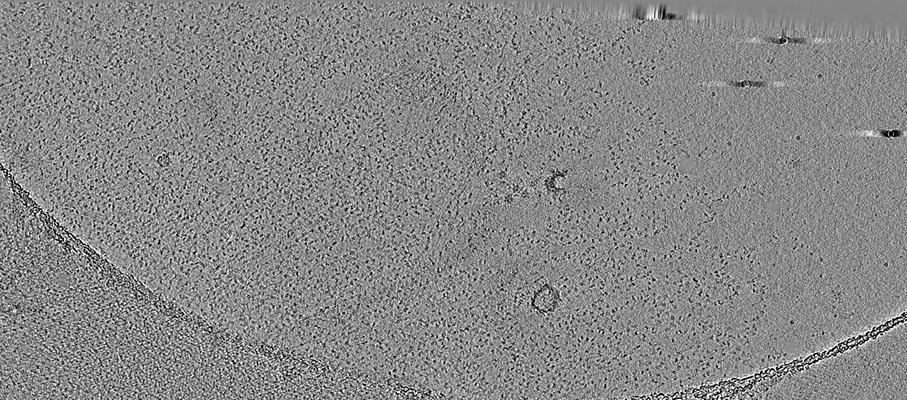
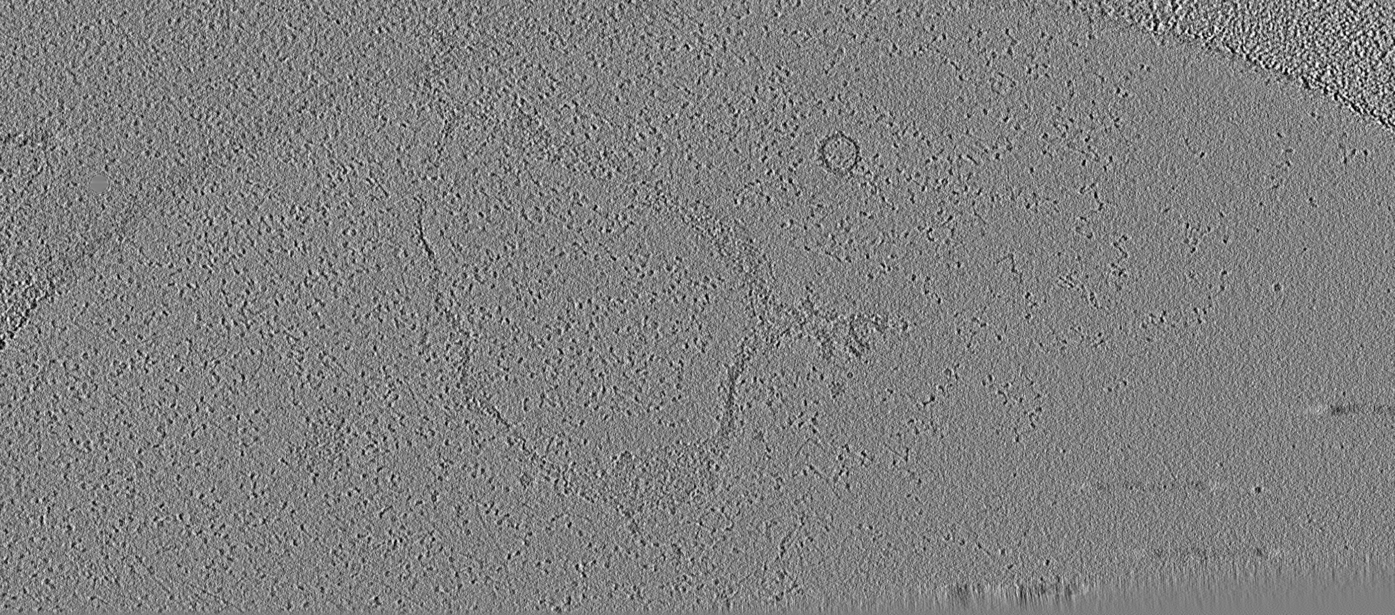
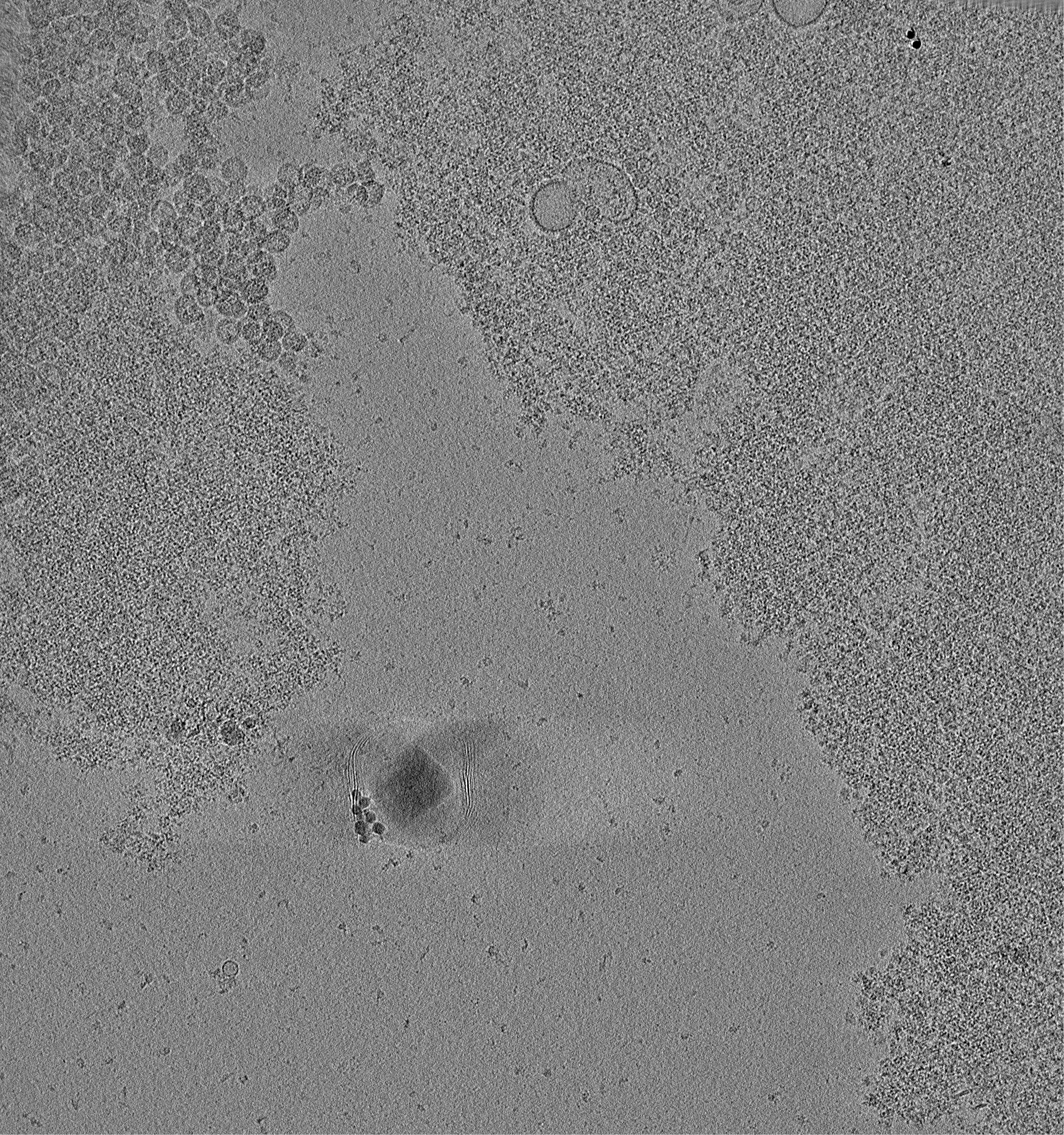
 Map data Map data | Cryotomogram of picoplankton chromatin in 5mM EDTA. | |||||||||
 Sample Sample |
| |||||||||
| Biological species |  Ostreococcus tauri (plant) Ostreococcus tauri (plant) | |||||||||
| Method | electron tomography / cryo EM | |||||||||
 Authors Authors | Cai S / Song Y / Chen C / Shi J / Gan L | |||||||||
 Citation Citation | Journal: Mol Biol Cell / Year: 2018 Title: Natural chromatin is heterogeneous and self-associates in vitro. Authors: Shujun Cai / Yajiao Song / Chen Chen / Jian Shi / Lu Gan /  Abstract: The 30-nm fiber is commonly formed by oligonucleosome arrays in vitro but rarely found inside cells. To determine how chromatin higher-order structure is controlled, we used electron cryotomography ...The 30-nm fiber is commonly formed by oligonucleosome arrays in vitro but rarely found inside cells. To determine how chromatin higher-order structure is controlled, we used electron cryotomography (cryo-ET) to study the undigested natural chromatin released from two single-celled organisms in which 30-nm fibers have not been observed in vivo: picoplankton and yeast. In the presence of divalent cations, most of the chromatin from both organisms is condensed into a large mass in vitro. Rare irregular 30-nm fibers, some of which include face-to-face nucleosome interactions, do form at the periphery of this mass. In the absence of divalent cations, picoplankton chromatin decondenses into open zigzags. By contrast, yeast chromatin mostly remains condensed, with very few open motifs. Yeast chromatin packing is largely unchanged in the absence of linker histone and mildly decondensed when histones are more acetylated. Natural chromatin is therefore generally nonpermissive of regular motifs, even at the level of oligonucleosomes. | |||||||||
| History |
|
- Structure visualization
| Movie |
 Movie viewer Movie viewer |
|---|---|
| Supplemental images |
- Downloads & links
-EMDB archive
| Map data | emd_6737.map.gz | 836.8 MB | EMDB map data format | |
|---|---|---|---|---|
| Header (meta data) | emd-6737-v30.xmlemd-6737.xml | 12.8 KB 12.8 KB | Display Display | EMDB header |
| Images |  emd_6737.png emd_6737.png | 700.4 KB | ||
| Others | emd_6737_additional.map.gz | 3.6 GB | ||
| Archive directory |  http://ftp.pdbj.org/pub/emdb/structures/EMD-6737ftp://ftp.pdbj.org/pub/emdb/structures/EMD-6737 http://ftp.pdbj.org/pub/emdb/structures/EMD-6737ftp://ftp.pdbj.org/pub/emdb/structures/EMD-6737 | HTTPS FTP |
-Related structure data
| EM raw data | EMPIAR-10098 (Title: Cryo-ET of natural chromatin from Ostreococcus tauri and Saccharyomyces cerevisiae Data size: 40.1 Data #1: Tilt series of chromatin release from picoplankton and budding yeast [class averages]) |
|---|
-Links
| EMDB pages | EMDB (EBI/PDBe) / EMDataResource |
|---|
-Map
| File | Download / File: emd_6737.map.gz / Format: CCP4 / Size: 1.1 GB / Type: IMAGE STORED AS SIGNED INTEGER (2 BYTES) | ||||||||||||||||||||||||||||||||||||||||||||||||||||||||||||
|---|---|---|---|---|---|---|---|---|---|---|---|---|---|---|---|---|---|---|---|---|---|---|---|---|---|---|---|---|---|---|---|---|---|---|---|---|---|---|---|---|---|---|---|---|---|---|---|---|---|---|---|---|---|---|---|---|---|---|---|---|---|
| Annotation | Cryotomogram of picoplankton chromatin in 5mM EDTA. | ||||||||||||||||||||||||||||||||||||||||||||||||||||||||||||
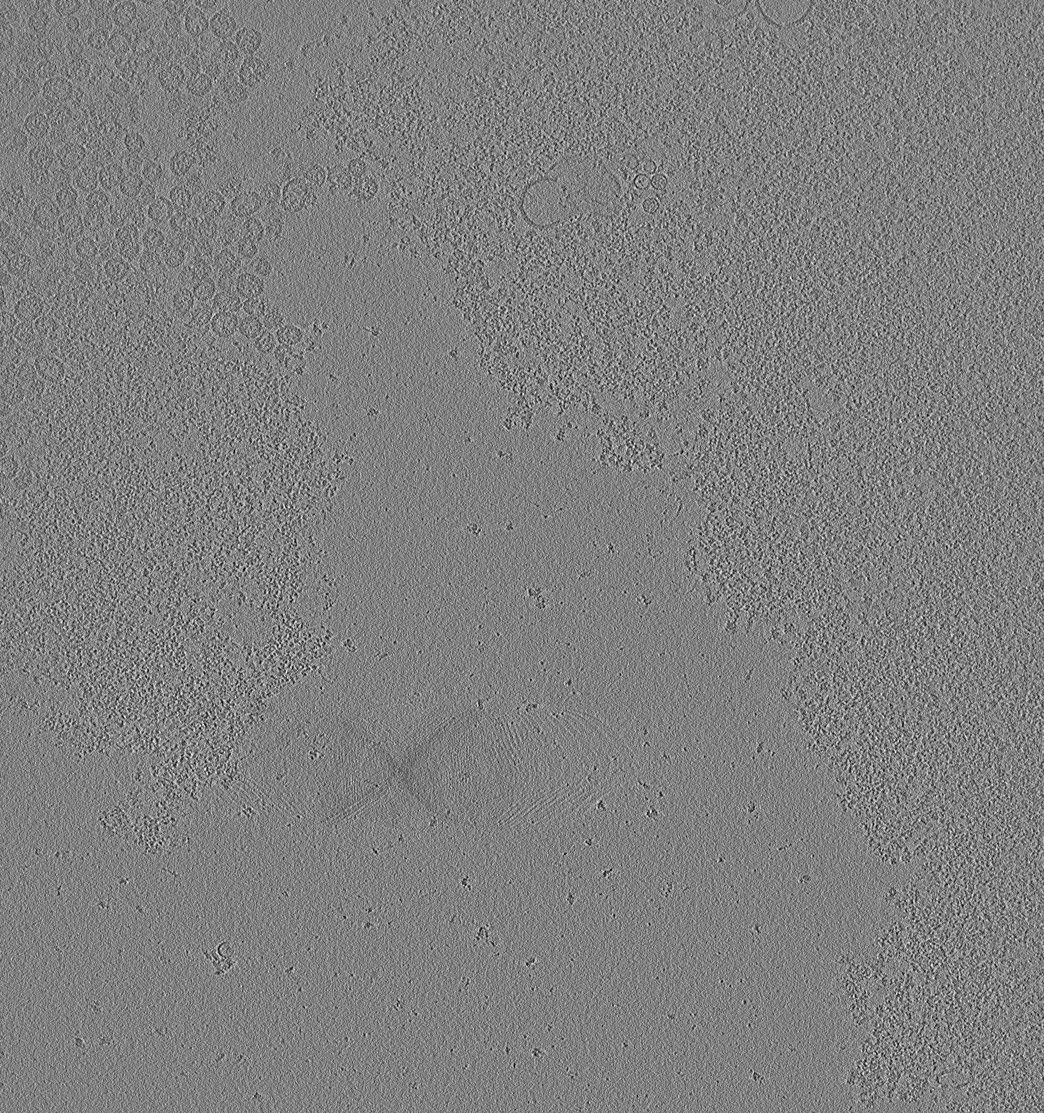
| Projections & slices | Image control
Images are generated by Spider. generated in cubic-lattice coordinate | ||||||||||||||||||||||||||||||||||||||||||||||||||||||||||||
| Voxel size | X=Y=Z: 9.12 Å | ||||||||||||||||||||||||||||||||||||||||||||||||||||||||||||
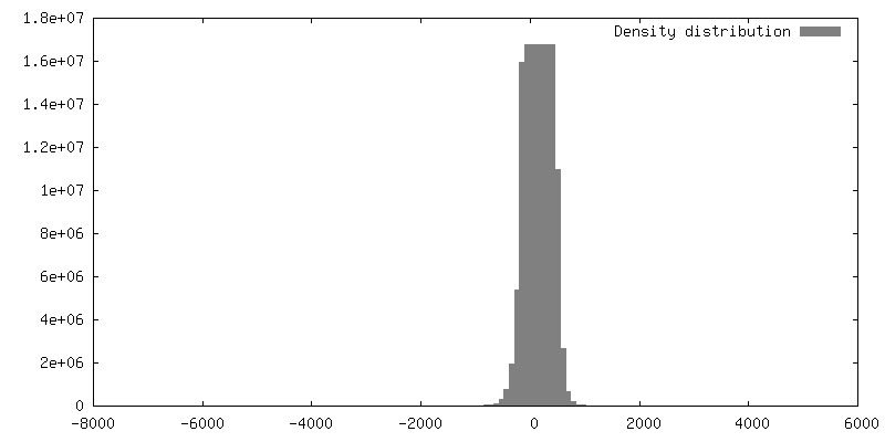
| Density |
| ||||||||||||||||||||||||||||||||||||||||||||||||||||||||||||
| Symmetry | Space group: 1 | ||||||||||||||||||||||||||||||||||||||||||||||||||||||||||||
| Details | EMDB XML:
CCP4 map header:
| ||||||||||||||||||||||||||||||||||||||||||||||||||||||||||||
 Z (Sec.)
 Y (Row.)
 X (Col.)


-Supplemental data
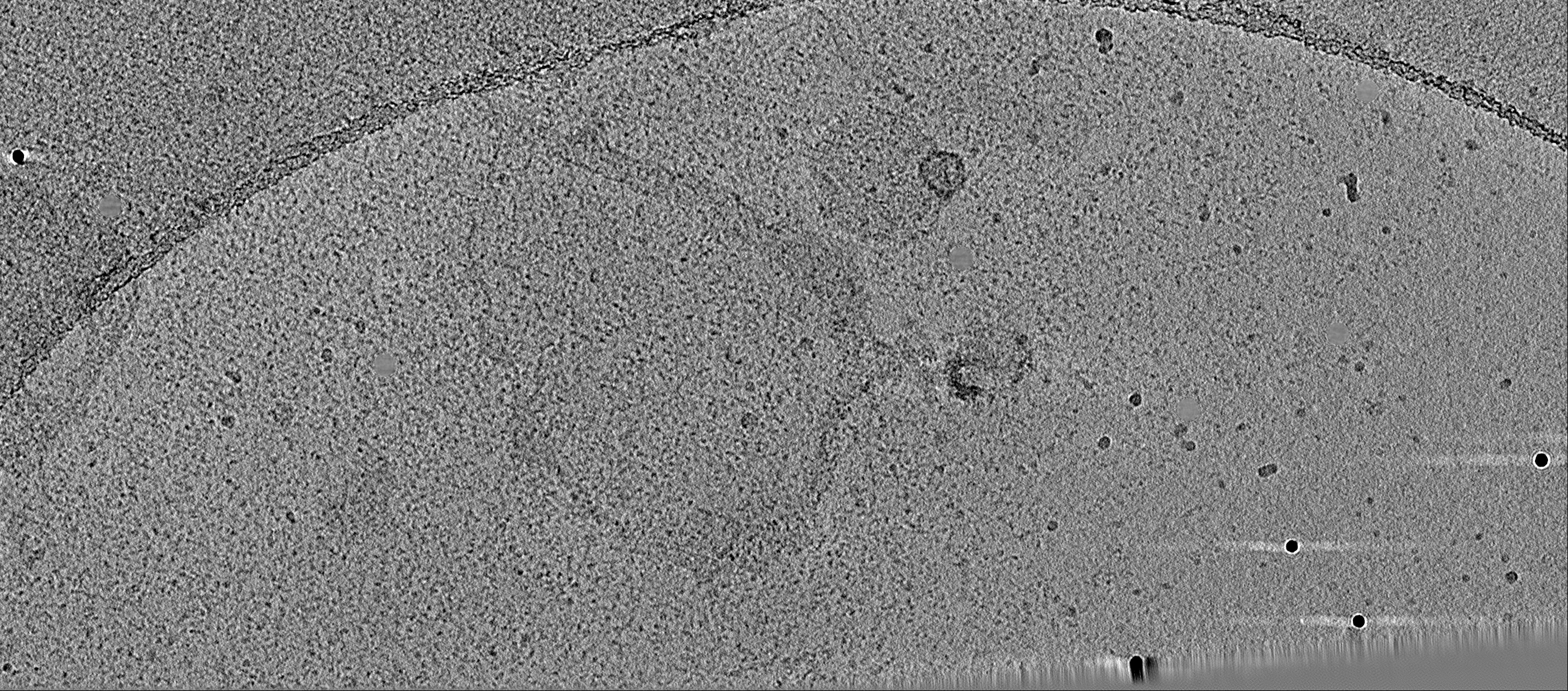
-Additional map: Cryotomogram of yeast chromatin in 50mM EDTA.
| File | emd_6737_additional.map | ||||||||||||
|---|---|---|---|---|---|---|---|---|---|---|---|---|---|
| Annotation | Cryotomogram of yeast chromatin in 50mM EDTA. | ||||||||||||
| Projections & Slices |
| ||||||||||||
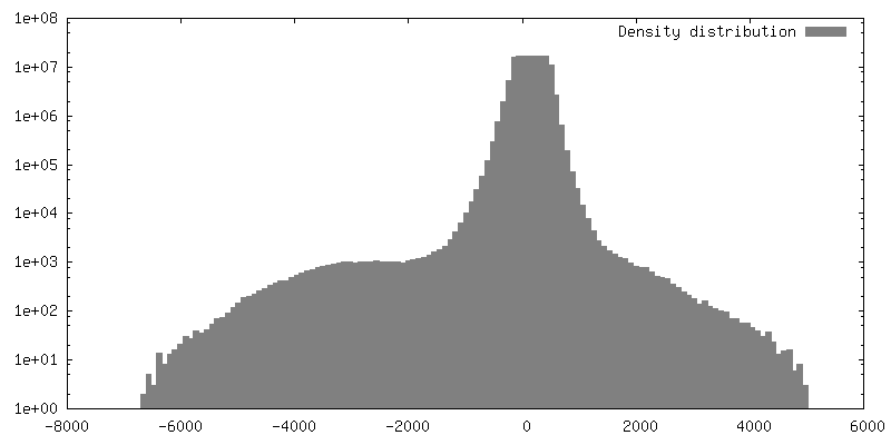
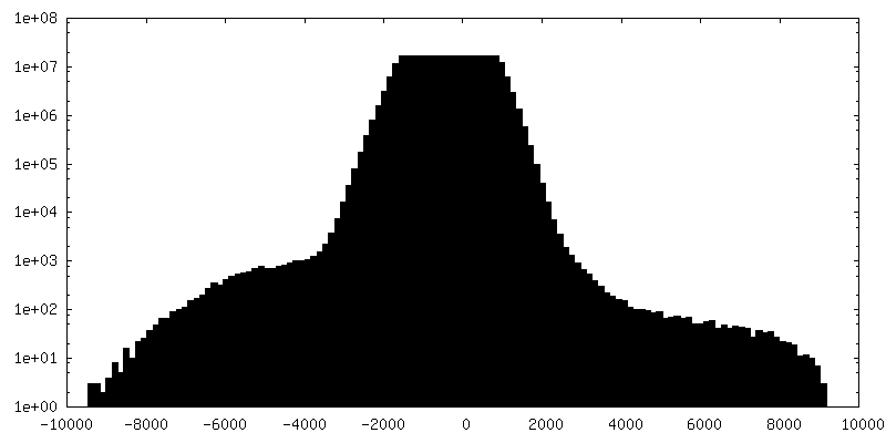
| Density Histograms |


- Sample components
-Entire : Chromatin released from the picoplankton Ostreococcus tauri, in 5...
| Entire | Name: Chromatin released from the picoplankton Ostreococcus tauri, in 5mM EDTA |
|---|---|
| Components |
|
-Supramolecule #1: Chromatin released from the picoplankton Ostreococcus tauri, in 5...
| Supramolecule | Name: Chromatin released from the picoplankton Ostreococcus tauri, in 5mM EDTA type: organelle_or_cellular_component / ID: 1 / Parent: 0 |
|---|---|
| Source (natural) | Organism: Ostreococcus tauri (plant) / Strain: RCC 745 |
-Experimental details
-Structure determination
| Method | cryo EM |
|---|---|
 Processing Processing | electron tomography |
| Aggregation state | filament |
-Sample preparation
| Buffer | pH: 7 Component:
| ||||||||||||
|---|---|---|---|---|---|---|---|---|---|---|---|---|---|
| Grid | Model: Protochips CF-4/2-2C-T / Material: COPPER / Mesh: 200 / Support film - Material: CARBON / Support film - topology: HOLEY / Pretreatment - Type: PLASMA CLEANING / Details: 15mA, Emitech K100X | ||||||||||||
| Vitrification | Cryogen name: NITROGEN / Chamber humidity: 100 % / Chamber temperature: 298 K / Instrument: FEI VITROBOT MARK IV | ||||||||||||
| Sectioning | Other: NO SECTIONING | ||||||||||||
| Fiducial marker | Manufacturer: Sigma / Diameter: 10 nm |
- Electron microscopy
| Microscope | FEI TITAN KRIOS |
|---|---|
| Image recording | Film or detector model: FEI FALCON II (4k x 4k) / Detector mode: INTEGRATING / Digitization - Dimensions - Width: 4096 pixel / Digitization - Dimensions - Height: 4096 pixel / Number grids imaged: 1 / Number real images: 62 / Average electron dose: 2.4 e/Å2 |
| Electron beam | Acceleration voltage: 300 kV / Electron source:  FIELD EMISSION GUN FIELD EMISSION GUN |
| Electron optics | Calibrated defocus min: 11.0 µm / Calibrated magnification: 15678 / Illumination mode: FLOOD BEAM / Imaging mode: BRIGHT FIELD / Cs: 2.7 mm / Nominal defocus min: 8.0 µm / Nominal magnification: 8700 |
| Sample stage | Specimen holder model: FEI TITAN KRIOS AUTOGRID HOLDER / Cooling holder cryogen: NITROGEN |
| Experimental equipment |  Model: Titan Krios / Image courtesy: FEI Company |
-Image processing
| Final reconstruction | Algorithm: BACK PROJECTION / Software - Name: IMOD (ver. 4.9) / Number images used: 59 |
|---|---|
| CTF correction | Software - Name: IMOD (ver. 4.8) |
