Journal: Cell / Year: 2017 Title: An Atomic Structure of the Human Spliceosome. Authors: Xiaofeng Zhang / Chuangye Yan / Jing Hang / Lorenzo I Finci / Jianlin Lei / Yigong Shi / Abstract: Mechanistic understanding of pre-mRNA splicing requires detailed structural information on various states of the spliceosome. Here we report the cryo electron microscopy (cryo-EM) structure of the ...Mechanistic understanding of pre-mRNA splicing requires detailed structural information on various states of the spliceosome. Here we report the cryo electron microscopy (cryo-EM) structure of the human spliceosome just before exon ligation (the C complex) at an average resolution of 3.76 Å. The splicing factor Prp17 stabilizes the active site conformation. The step II factor Slu7 adopts an extended conformation, binds Prp8 and Cwc22, and is poised for selection of the 3'-splice site. Remarkably, the intron lariat traverses through a positively charged central channel of RBM22; this unusual organization suggests mechanisms of intron recruitment, confinement, and release. The protein PRKRIP1 forms a 100-Å α helix linking the distant U2 snRNP to the catalytic center. A 35-residue fragment of the ATPase/helicase Prp22 latches onto Prp8, and the quaternary exon junction complex (EJC) recognizes upstream 5'-exon sequences and associates with Cwc22 and the GTPase Snu114. These structural features reveal important mechanistic insights into exon ligation.
History
Deposition
Apr 30, 2017
-
Header (metadata) release
Jun 28, 2017
-
Map release
Jul 5, 2017
-
Update
Nov 20, 2024
-
Current status
Nov 20, 2024
Processing site: PDBj / Status: Released
-
Structure visualization
Movie
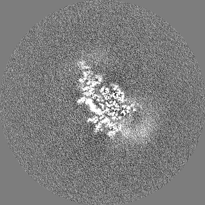
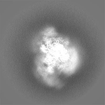
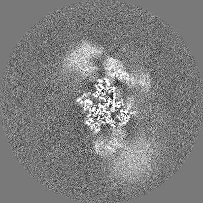
Surface view with section colored by density value
Chromatic aberration corrector: -10 / Energy filter - Name: GIF Quantum LS / Energy filter - Lower energy threshold: 0 eV / Energy filter - Upper energy threshold: 10 eV
Image recording
Film or detector model: GATAN K2 SUMMIT (4k x 4k) / Detector mode: SUPER-RESOLUTION / Digitization - Frames/image: 2-32 / Number grids imaged: 5 / Number real images: 7308 / Average exposure time: 0.25 sec. / Average electron dose: 50.0 e/Å2
Electron beam
Acceleration voltage: 300 kV / Electron source: FIELD EMISSION GUN
Electron optics
Illumination mode: FLOOD BEAM / Imaging mode: BRIGHT FIELD / Cs: 2.7 mm
In the structure databanks used in Yorodumi, some data are registered as the other names, "COVID-19 virus" and "2019-nCoV". Here are the details of the virus and the list of structure data.
Jan 31, 2019. EMDB accession codes are about to change! (news from PDBe EMDB page)
EMDB accession codes are about to change! (news from PDBe EMDB page)
The allocation of 4 digits for EMDB accession codes will soon come to an end. Whilst these codes will remain in use, new EMDB accession codes will include an additional digit and will expand incrementally as the available range of codes is exhausted. The current 4-digit format prefixed with “EMD-” (i.e. EMD-XXXX) will advance to a 5-digit format (i.e. EMD-XXXXX), and so on. It is currently estimated that the 4-digit codes will be depleted around Spring 2019, at which point the 5-digit format will come into force.
The EM Navigator/Yorodumi systems omit the EMD- prefix.
Related info.:Q: What is EMD? / ID/Accession-code notation in Yorodumi/EM Navigator
Yorodumi is a browser for structure data from EMDB, PDB, SASBDB, etc.
This page is also the successor to EM Navigator detail page, and also detail information page/front-end page for Omokage search.
The word "yorodu" (or yorozu) is an old Japanese word meaning "ten thousand". "mi" (miru) is to see.
Related info.:EMDB / PDB / SASBDB / Comparison of 3 databanks / Yorodumi Search / Aug 31, 2016. New EM Navigator & Yorodumi / Yorodumi Papers / Jmol/JSmol / Function and homology information / Changes in new EM Navigator and Yorodumi
 Movie
Movie Controller

 Yorodumi
 Open data
 Basic information
 Map data
 Sample
 Keywords
 Function and homology information
 Homo sapiens (human) /
 Human adenovirus 2
 Authors
 China, 2 items
  Citation
 Structure visualization
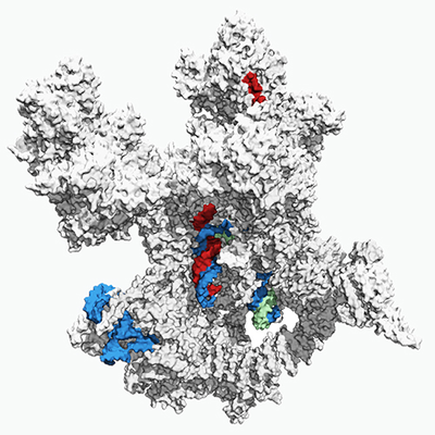
 Downloads & links
 emd_6721.png
 http://ftp.pdbj.org/pub/emdb/structures/EMD-6721


 Z (Sec.)
 Y (Row.)
 X (Col.)


 Sample components


 Processing
 Electron microscopy
 FIELD EMISSION GUN

