 Movie
Movie Controller


+ Open data

- Basic information
| Entry | Database: EMDB / ID: EMD-6669 | |||||||||
|---|---|---|---|---|---|---|---|---|---|---|
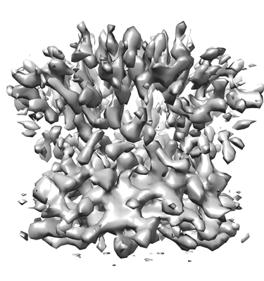
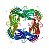
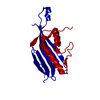
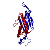
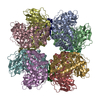
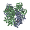
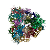
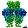
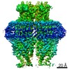
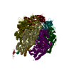
| Title | The endolysosomal Ca2+ channel TRPML1 | |||||||||
 Map data Map data | TrpML1 density map at 8.12A resolution, sharpened by post-processing procedure in RELION | |||||||||
 Sample Sample |
| |||||||||
| Biological species |   Caenorhabditis elegans (invertebrata) Caenorhabditis elegans (invertebrata) | |||||||||
| Method | single particle reconstruction / cryo EM / Resolution: 8.12 Å | |||||||||
 Authors Authors | Li X / Zhou X | |||||||||
 Citation Citation | Journal: Nat Struct Mol Biol / Year: 2017 Title: Structural basis of dual Ca/pH regulation of the endolysosomal TRPML1 channel. Authors: Minghui Li / Wei K Zhang / Nicole M Benvin / Xiaoyuan Zhou / Deyuan Su / Huan Li / Shu Wang / Ioannis E Michailidis / Liang Tong / Xueming Li / Jian Yang /   Abstract: The activities of organellar ion channels are often regulated by Ca and H, which are present in high concentrations in many organelles. Here we report a structural element critical for dual Ca/pH ...The activities of organellar ion channels are often regulated by Ca and H, which are present in high concentrations in many organelles. Here we report a structural element critical for dual Ca/pH regulation of TRPML1, a Ca-release channel crucial for endolysosomal function. TRPML1 mutations cause mucolipidosis type IV (MLIV), a severe lysosomal storage disorder characterized by neurodegeneration, mental retardation and blindness. We obtained crystal structures of the 213-residue luminal domain of human TRPML1 containing three missense MLIV-causing mutations. This domain forms a tetramer with a highly electronegative central pore formed by a novel luminal pore loop. Cysteine cross-linking and cryo-EM analyses confirmed that this architecture occurs in the full-length channel. Structure-function studies demonstrated that Ca and H interact with the luminal pore and exert physiologically important regulation. The MLIV-causing mutations disrupt the luminal-domain structure and cause TRPML1 mislocalization. Our study reveals the structural underpinnings of TRPML1's regulation, assembly and pathogenesis. | |||||||||
| History |
|
- Structure visualization
| Movie |
 Movie viewer Movie viewer |
|---|---|
| Structure viewer | EM map: SurfViewMolmilJmol/JSmol |
| Supplemental images |
- Downloads & links
-EMDB archive
| Map data | emd_6669.map.gz | 348.3 KB | EMDB map data format | |
|---|---|---|---|---|
| Header (meta data) | emd-6669-v30.xmlemd-6669.xml | 16 KB 16 KB | Display Display | EMDB header |
| Images |  emd_6669.png emd_6669.png | 83.6 KB | ||
| Others | emd_6669_additional.map.gz | 981.1 KB | ||
| Archive directory |  http://ftp.pdbj.org/pub/emdb/structures/EMD-6669ftp://ftp.pdbj.org/pub/emdb/structures/EMD-6669 http://ftp.pdbj.org/pub/emdb/structures/EMD-6669ftp://ftp.pdbj.org/pub/emdb/structures/EMD-6669 | HTTPS FTP |
-Related structure data


-Links
| EMDB pages | EMDB (EBI/PDBe) / EMDataResource |
|---|
-Map
| File | Download / File: emd_6669.map.gz / Format: CCP4 / Size: 2 MB / Type: IMAGE STORED AS FLOATING POINT NUMBER (4 BYTES) | ||||||||||||||||||||||||||||||||||||||||||||||||||||||||||||
|---|---|---|---|---|---|---|---|---|---|---|---|---|---|---|---|---|---|---|---|---|---|---|---|---|---|---|---|---|---|---|---|---|---|---|---|---|---|---|---|---|---|---|---|---|---|---|---|---|---|---|---|---|---|---|---|---|---|---|---|---|---|
| Annotation | TrpML1 density map at 8.12A resolution, sharpened by post-processing procedure in RELION | ||||||||||||||||||||||||||||||||||||||||||||||||||||||||||||
| Voxel size | X=Y=Z: 2.64 Å | ||||||||||||||||||||||||||||||||||||||||||||||||||||||||||||
| Density |
| ||||||||||||||||||||||||||||||||||||||||||||||||||||||||||||
| Symmetry | Space group: 1 | ||||||||||||||||||||||||||||||||||||||||||||||||||||||||||||
| Details | EMDB XML:
CCP4 map header:
| ||||||||||||||||||||||||||||||||||||||||||||||||||||||||||||
-Supplemental data
-Additional map: TrpML1 density map, not sharpened
| File | emd_6669_additional.map | ||||||||||||
|---|---|---|---|---|---|---|---|---|---|---|---|---|---|
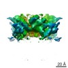
| Annotation | TrpML1 density map, not sharpened | ||||||||||||
| Projections & Slices |
| ||||||||||||
| Density Histograms |
 Z
Z Y
Y X
X


- Sample components
-Entire : The endolysosomal Ca2+ channel TRPML1
| Entire | Name: The endolysosomal Ca2+ channel TRPML1 |
|---|---|
| Components |
|
-Supramolecule #1: The endolysosomal Ca2+ channel TRPML1
| Supramolecule | Name: The endolysosomal Ca2+ channel TRPML1 / type: complex / ID: 1 / Parent: 0 / Macromolecule list: all |
|---|---|
| Recombinant expression | Organism:  Trichoplusia ni (cabbage looper) / Recombinant plasmid: pFastBac1 Trichoplusia ni (cabbage looper) / Recombinant plasmid: pFastBac1 |
-Macromolecule #1: The endolysosomal Ca2+ channel TRPML1
| Macromolecule | Name: The endolysosomal Ca2+ channel TRPML1 / type: protein_or_peptide / ID: 1 / Enantiomer: LEVO |
|---|---|
| Source (natural) | Organism: Caenorhabditis elegans (invertebrata) |
| Recombinant expression | Organism: Trichoplusia ni (cabbage looper) |
| Sequence | String: GGGGSMSRRS TTDRSDFNDN ASNASSNASR RPTINFQEIM EDLPHHERET GERLRRHLQF FFMNPMEKWK VRHQLPYKLV LQVLKIVFVT MQLILFAEMR MSHVDFLEDT TTVMRHRFLK EWNDDRDALQ YPPAEGRYSV YDDQGLSEHL SFLINSYYSI RNDSFASFSY ...String: GGGGSMSRRS TTDRSDFNDN ASNASSNASR RPTINFQEIM EDLPHHERET GERLRRHLQF FFMNPMEKWK VRHQLPYKLV LQVLKIVFVT MQLILFAEMR MSHVDFLEDT TTVMRHRFLK EWNDDRDALQ YPPAEGRYSV YDDQGLSEHL SFLINSYYSI RNDSFASFSY DVVSHPSGNL GAQISFESIP PIEVLIDRIS NVTVNNNTYN FDIREVKDTK RLNLTETEVF QIGQSDDAVR DILATRGITF LPEDALKIST VQFKFRLRTI HYSPTAGDQK PECYKISVSI KFDNSRHTGQ VHVTLSTVVS YVNVCNGRII KGVGWSFDTL LIGGTDIFVL ILCILSLILC CRALIKAHLL QIKTSDYFEN VLKNKITVTD QLDFLNLWYV MIVVNDALII IGTVAKISIE FQDFDNSLFT LTSIFLGMGA LLVYVGVLRY FGFFSQYNIL MLTLKRSAPN IMRFMTCAIV LYAGFLIAGW VIIGPYSMKF RTLAESSEAL FSLLNGDDMF ATFYTINDSN TVIKVFGTVY IYLFVSLFIY VVLSLFIAII MDAYEVVKDR YSDGLRAIEK RGCLRDFVES NPPPSELGSP TTRSAYAPSN LLNLGRGWQR LE |
-Experimental details
-Structure determination
| Method | cryo EM |
|---|---|
 Processing Processing | single particle reconstruction |
| Aggregation state | particle |
-Sample preparation
| Concentration | 0.6 mg/mL | |||||||||
|---|---|---|---|---|---|---|---|---|---|---|
| Buffer | pH: 7.4 Component:
| |||||||||
| Grid | Model: R1.2/1.3 / Material: COPPER / Mesh: 400 / Support film - Material: CARBON / Support film - topology: HOLEY ARRAY / Pretreatment - Type: GLOW DISCHARGE / Pretreatment - Atmosphere: AIR / Pretreatment - Pressure: 0.039 kPa | |||||||||
| Vitrification | Cryogen name: ETHANE / Chamber humidity: 100 % / Chamber temperature: 281.15 K / Instrument: FEI VITROBOT MARK IV Details: waiting for 3 seconds before blotting for 3.5 seconds(double-sided,blot force 1), then the grid was immediately plunged into liquid ethane cooled by liquid-nitrogen.. | |||||||||
| Details | the sample was homogeneous |
- Electron microscopy
| Microscope | FEI TITAN KRIOS |
|---|---|
| Electron beam | Acceleration voltage: 300 kV / Electron source: FIELD EMISSION GUN |
| Electron optics | Illumination mode: SPOT SCAN / Imaging mode: BRIGHT FIELDBright-field microscopy / Cs: 2.7 mm / Nominal defocus max: 3.1 µm / Nominal defocus min: 2.1 µm / Nominal magnification: 22500 |
| Sample stage | Specimen holder model: FEI TITAN KRIOS AUTOGRID HOLDER / Cooling holder cryogen: NITROGEN |
| Image recording | Film or detector model: GATAN K2 SUMMIT (4k x 4k) / Detector mode: SUPER-RESOLUTION / Digitization - Dimensions - Width: 7676 pixel / Digitization - Dimensions - Height: 7420 pixel / Digitization - Sampling interval: 5.0 µm / Digitization - Frames/image: 1-32 / Average exposure time: 8.0 sec. / Average electron dose: 50.0 e/Å2 |
| Experimental equipment |  Model: Titan Krios / Image courtesy: FEI Company |
-Image processing
| CTF correction | Software - Name: CTFFIND (ver. 3) |
|---|---|
| Startup model | Type of model: OTHER Details: an four pieces of propeller was drawn manually as initial model |
| Initial angle assignment | Type: PROJECTION MATCHING / Software - Name: RELION (ver. 1.4) |
| Final 3D classification | Software - Name: RELION (ver. 1.4) |
| Final angle assignment | Type: PROJECTION MATCHING / Software - Name: RELION (ver. 1.4) |
| Final reconstruction | Applied symmetry - Point group: C4 (4 fold cyclic) / Resolution.type: BY AUTHOR / Resolution: 8.12 Å / Resolution method: FSC 0.143 CUT-OFF / Software - Name: RELION (ver. 1.4) / Number images used: 71052 |
