 Movie
Movie Controller

[English] 日本語
 Yorodumi
- EMDB-6558: Cryo-EM of bacteriophage phi29 emptied particles fusing with lipo... -
+ Open data

- Basic information
| Entry | Database: EMDB / ID: EMD-6558 | |||||||||
|---|---|---|---|---|---|---|---|---|---|---|
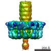
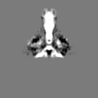
| Title | Cryo-EM of bacteriophage phi29 emptied particles fusing with liposome after low pH treatment | |||||||||
 Map data Map data | reconstruction of bacteriophage phi29 fusing with liposomes after low pH treatment | |||||||||
 Sample Sample |
| |||||||||
 Keywords Keywords | bacteriophage phi29 / empty particles / tail knob protein gp9 | |||||||||
| Biological species |   Bacillus phage phi29 (virus) Bacillus phage phi29 (virus) | |||||||||
| Method | single particle reconstruction / cryo EM / Resolution: 34.5 Å | |||||||||
 Authors Authors | Xu JW / Gui M / Wang DH / Xiang Y | |||||||||
 Citation Citation | Journal: Nature / Year: 2016 Title: The bacteriophage ϕ29 tail possesses a pore-forming loop for cell membrane penetration. Authors: Jingwei Xu / Miao Gui / Dianhong Wang / Ye Xiang /  Abstract: Most bacteriophages are tailed bacteriophages with an isometric or a prolate head attached to a long contractile, long non-contractile, or short non-contractile tail. The tail is a complex machine ...Most bacteriophages are tailed bacteriophages with an isometric or a prolate head attached to a long contractile, long non-contractile, or short non-contractile tail. The tail is a complex machine that plays a central role in host cell recognition and attachment, cell wall and membrane penetration, and viral genome ejection. The mechanisms involved in the penetration of the inner host cell membrane by bacteriophage tails are not well understood. Here we describe structural and functional studies of the bacteriophage ϕ29 tail knob protein gene product 9 (gp9). The 2.0 Å crystal structure of gp9 shows that six gp9 molecules form a hexameric tube structure with six flexible hydrophobic loops blocking one end of the tube before DNA ejection. Sequence and structural analyses suggest that the loops in the tube could be membrane active. Further biochemical assays and electron microscopy structural analyses show that the six hydrophobic loops in the tube exit upon DNA ejection and form a channel that spans the lipid bilayer of the membrane and allows the release of the bacteriophage genomic DNA, suggesting that cell membrane penetration involves a pore-forming mechanism similar to that of certain non-enveloped eukaryotic viruses. A search of other phage tail proteins identified similar hydrophobic loops, which indicates that a common mechanism might be used for membrane penetration by prokaryotic viruses. These findings suggest that although prokaryotic and eukaryotic viruses use apparently very different mechanisms for infection, they have evolved similar mechanisms for breaching the cell membrane. | |||||||||
| History |
|
- Structure visualization
| Movie |
 Movie viewer Movie viewer |
|---|---|
| Structure viewer | EM map: SurfViewMolmilJmol/JSmol |
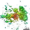
| Supplemental images |

- Downloads & links
-EMDB archive
| Map data | emd_6558.map.gz | 7.8 MB | EMDB map data format | |
|---|---|---|---|---|
| Header (meta data) | emd-6558-v30.xmlemd-6558.xml | 8 KB 8 KB | Display Display | EMDB header |
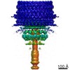
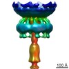
| Images | 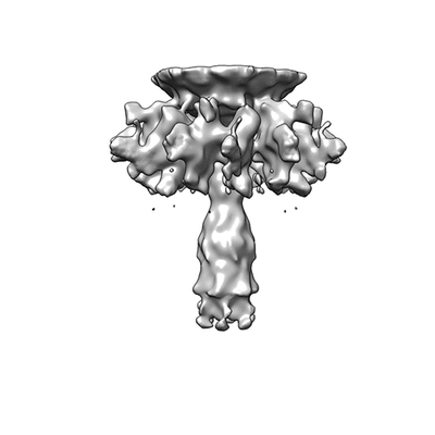 400_6558.gif 400_6558.gif 80_6558.gif 80_6558.gif | 36.4 KB 14.9 KB | ||
| Archive directory |  http://ftp.pdbj.org/pub/emdb/structures/EMD-6558ftp://ftp.pdbj.org/pub/emdb/structures/EMD-6558 http://ftp.pdbj.org/pub/emdb/structures/EMD-6558ftp://ftp.pdbj.org/pub/emdb/structures/EMD-6558 | HTTPS FTP |
-Related structure data


-Links
| EMDB pages | EMDB (EBI/PDBe) / EMDataResource |
|---|
-Map
| File | Download / File: emd_6558.map.gz / Format: CCP4 / Size: 122.1 MB / Type: IMAGE STORED AS FLOATING POINT NUMBER (4 BYTES) | ||||||||||||||||||||||||||||||||||||||||||||||||||||||||||||||||||||
|---|---|---|---|---|---|---|---|---|---|---|---|---|---|---|---|---|---|---|---|---|---|---|---|---|---|---|---|---|---|---|---|---|---|---|---|---|---|---|---|---|---|---|---|---|---|---|---|---|---|---|---|---|---|---|---|---|---|---|---|---|---|---|---|---|---|---|---|---|---|
| Annotation | reconstruction of bacteriophage phi29 fusing with liposomes after low pH treatment | ||||||||||||||||||||||||||||||||||||||||||||||||||||||||||||||||||||
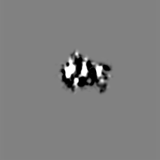
| Projections & slices | Image control
Images are generated by Spider. | ||||||||||||||||||||||||||||||||||||||||||||||||||||||||||||||||||||
| Voxel size | X=Y=Z: 3.02 Å | ||||||||||||||||||||||||||||||||||||||||||||||||||||||||||||||||||||
| Density |
| ||||||||||||||||||||||||||||||||||||||||||||||||||||||||||||||||||||
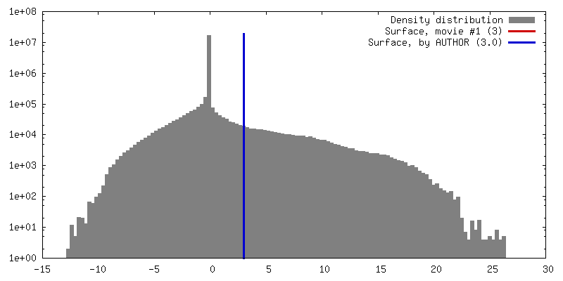
| Symmetry | Space group: 1 | ||||||||||||||||||||||||||||||||||||||||||||||||||||||||||||||||||||
| Details | EMDB XML:
CCP4 map header:
| ||||||||||||||||||||||||||||||||||||||||||||||||||||||||||||||||||||
 Z (Sec.)
 Y (Row.)
 X (Col.)


-Supplemental data
- Sample components
-Entire : the tail of bacteriophage phi29 fusing with liposomes after low p...
| Entire | Name: the tail of bacteriophage phi29 fusing with liposomes after low pH treatment |
|---|---|
| Components |
|
-Supramolecule #1000: the tail of bacteriophage phi29 fusing with liposomes after low p...
| Supramolecule | Name: the tail of bacteriophage phi29 fusing with liposomes after low pH treatment type: sample / ID: 1000 / Oligomeric state: 1 / Number unique components: 1 |
|---|
-Supramolecule #1: Bacillus phage phi29
| Supramolecule | Name: Bacillus phage phi29 / type: virus / ID: 1 / NCBI-ID: 10756 / Sci species name: Bacillus phage phi29 / Database: NCBI / Virus type: VIRION / Virus isolate: SPECIES / Virus enveloped: No / Virus empty: Yes |
|---|---|
| Host (natural) | Organism:  |
| Virus shell | Shell ID: 1 / Name: capsid protein, gp8 |
-Experimental details
-Structure determination
| Method | cryo EM |
|---|---|
 Processing Processing | single particle reconstruction |
| Aggregation state | particle |
-Sample preparation
| Buffer | pH: 4.2 Details: phage in the 20mM NaCl, 10mM Tris-Cl, 10mM MgCl2 mixed with 50mM NaAc, 150mM (NH4)2SO4, 12.5mM HEPES, 50mM KCl solution |
|---|---|
| Grid | Details: Quantifoild 200 mesh grid, 1.2 um x 1.3 um |
| Vitrification | Cryogen name: ETHANE / Chamber humidity: 90 % / Instrument: GATAN CRYOPLUNGE 3 |
- Electron microscopy
| Microscope | FEI TECNAI F20 |
|---|---|
| Date | Apr 7, 2015 |
| Image recording | Category: CCD / Film or detector model: FEI EAGLE (4k x 4k) / Number real images: 191 / Average electron dose: 20 e/Å2 |
| Electron beam | Acceleration voltage: 200 kV / Electron source:  FIELD EMISSION GUN FIELD EMISSION GUN |
| Electron optics | Illumination mode: SPOT SCAN / Imaging mode: BRIGHT FIELD / Cs: 2.7 mm |
| Sample stage | Specimen holder: 626 holder / Specimen holder model: GATAN HELIUM |
| Experimental equipment |  Model: Tecnai F20 / Image courtesy: FEI Company |
-Image processing
| CTF correction | Details: Each particle |
|---|---|
| Final reconstruction | Resolution.type: BY AUTHOR / Resolution: 34.5 Å / Resolution method: OTHER / Software - Name: EMAN2 / Number images used: 802 |
