 Movie
Movie Controller


+ Open data

- Basic information
| Entry | Database: EMDB / ID: EMD-6554 | |||||||||
|---|---|---|---|---|---|---|---|---|---|---|
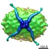
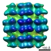
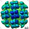
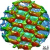
| Title | Cryo-Molecular electron tomography of IgM | |||||||||
 Map data Map data | IgM extended | |||||||||
 Sample Sample |
| |||||||||
 Keywords Keywords | Malaria / Rosetting / IgM | |||||||||
| Biological species |  Homo sapiens (human) Homo sapiens (human) | |||||||||
| Method | electron tomography / cryo EM / Resolution: 25.0 Å | |||||||||
 Authors Authors | Akhouri RR / Goel S / Furusho H / Skoglund U / Wahlgren M | |||||||||
 Citation Citation | Journal: Cell Rep / Year: 2016 Title: Architecture of Human IgM in Complex with P. falciparum Erythrocyte Membrane Protein 1. Authors: Reetesh Raj Akhouri / Suchi Goel / Hirotoshi Furusho / Ulf Skoglund / Mats Wahlgren /   Abstract: Plasmodium falciparum virulence is associated with sequestration of infected erythrocytes. Microvascular binding mediated by PfEMP1 in complex with non-immune immunoglobulin M (IgM) is common among ...Plasmodium falciparum virulence is associated with sequestration of infected erythrocytes. Microvascular binding mediated by PfEMP1 in complex with non-immune immunoglobulin M (IgM) is common among parasites that cause both severe childhood malaria and pregnancy-associated malaria. Here, we present cryo-molecular electron tomography structures of human IgM, PfEMP1 and their complex. Three-dimensional reconstructions of IgM reveal that it has a dome-like core, randomly oriented Fab2s units, and the overall shape of a turtle. PfEMP1 is a C- shaped molecule with a flexible N terminus followed by an arc-shaped backbone and a bulky C terminus that interacts with IgM. Our data demonstrate that the PfEMP1 binding pockets on IgM overlap with those of C1q, and the bulkiness of PfEMP1 limits the capacity of IgM to interact with PfEMP1. We suggest that P. falciparum exploits IgM to cluster PfEMP1 into an organized matrix to augment its affinity to host cell receptors. | |||||||||
| History |
|
- Structure visualization
| Movie |
 Movie viewer Movie viewer |
|---|---|
| Structure viewer | EM map: SurfViewMolmilJmol/JSmol |
| Supplemental images |

- Downloads & links
-EMDB archive
| Map data | emd_6554.map.gz | 7.8 MB | EMDB map data format | |
|---|---|---|---|---|
| Header (meta data) | emd-6554-v30.xmlemd-6554.xml | 9.5 KB 9.5 KB | Display Display | EMDB header |
| Images | emd_6554.tif | 699.6 KB | ||
| Archive directory |  http://ftp.pdbj.org/pub/emdb/structures/EMD-6554ftp://ftp.pdbj.org/pub/emdb/structures/EMD-6554 http://ftp.pdbj.org/pub/emdb/structures/EMD-6554ftp://ftp.pdbj.org/pub/emdb/structures/EMD-6554 | HTTPS FTP |
-Related structure data
| Related structure data | 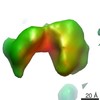 2882C  2883C 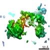 2884C 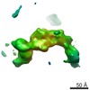 2885C 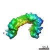 2886C 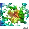 2887C 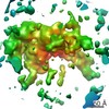 2888C 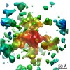 2889C 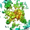 2890C 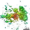 2891C 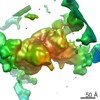 2892C  2893C 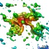 2894C 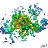 2895C C: citing same article ( |
|---|---|
| Similar structure data |
-Links
| EMDB pages | EMDB (EBI/PDBe) / EMDataResource |
|---|
-Map
| File | Download / File: emd_6554.map.gz / Format: CCP4 / Size: 8.7 MB / Type: IMAGE STORED AS FLOATING POINT NUMBER (4 BYTES) | ||||||||||||||||||||||||||||||||||||||||||||||||||||||||||||
|---|---|---|---|---|---|---|---|---|---|---|---|---|---|---|---|---|---|---|---|---|---|---|---|---|---|---|---|---|---|---|---|---|---|---|---|---|---|---|---|---|---|---|---|---|---|---|---|---|---|---|---|---|---|---|---|---|---|---|---|---|---|
| Annotation | IgM extended | ||||||||||||||||||||||||||||||||||||||||||||||||||||||||||||
| Projections & slices | Image control
Images are generated by Spider. generated in cubic-lattice coordinate | ||||||||||||||||||||||||||||||||||||||||||||||||||||||||||||
| Voxel size | X=Y=Z: 2.267 Å | ||||||||||||||||||||||||||||||||||||||||||||||||||||||||||||
| Density |
| ||||||||||||||||||||||||||||||||||||||||||||||||||||||||||||
| Symmetry | Space group: 1 | ||||||||||||||||||||||||||||||||||||||||||||||||||||||||||||
| Details | EMDB XML:
CCP4 map header:
| ||||||||||||||||||||||||||||||||||||||||||||||||||||||||||||
 Z (Sec.)
 Y (Row.)
 X (Col.)


-Supplemental data
- Sample components
-Entire : IgM extended
| Entire | Name: IgM extended |
|---|---|
| Components |
|
-Supramolecule #1000: IgM extended
| Supramolecule | Name: IgM extended / type: sample / ID: 1000 / Oligomeric state: monomer / Number unique components: 1 |
|---|---|
| Molecular weight | Experimental: 950 KDa / Theoretical: 950 KDa |
-Macromolecule #1: Immunoglobulin M
| Macromolecule | Name: Immunoglobulin M / type: protein_or_peptide / ID: 1 / Name.synonym: IgM / Number of copies: 1 / Oligomeric state: monomer / Recombinant expression: No / Database: NCBI |
|---|---|
| Source (natural) | Organism: Homo sapiens (human) / synonym: human |
| Molecular weight | Experimental: 950 KDa / Theoretical: 950 KDa |
-Experimental details
-Structure determination
| Method | cryo EM |
|---|---|
 Processing Processing | electron tomography |
| Aggregation state | particle |
-Sample preparation
| Concentration | 2.0 mg/mL |
|---|---|
| Buffer | pH: 7.5 / Details: 20 mM HEPES, 200 mM NaCl |
| Grid | Details: C-Flat (Copper grid with thin Carbon support) |
| Vitrification | Cryogen name: ETHANE / Chamber humidity: 100 % / Chamber temperature: 78 K / Instrument: FEI VITROBOT MARK IV / Method: Blot for 4 seconds before plunging. |
- Electron microscopy
| Microscope | FEI TITAN KRIOS |
|---|---|
| Temperature | Min: 78 K / Max: 100 K |
| Specialist optics | Energy filter - Name: FEI |
| Date | Jun 13, 2014 |
| Image recording | Category: CCD / Film or detector model: FEI FALCON II (4k x 4k) / Number real images: 281 / Average electron dose: 40 e/Å2 |
| Electron beam | Acceleration voltage: 300 kV / Electron source:  FIELD EMISSION GUN FIELD EMISSION GUN |
| Electron optics | Illumination mode: OTHER / Imaging mode: BRIGHT FIELD / Cs: 2.7 mm / Nominal defocus max: 1.5 µm / Nominal defocus min: 0.8 µm / Nominal magnification: 37000 |
| Sample stage | Specimen holder: LN2-cooled / Specimen holder model: FEI TITAN KRIOS AUTOGRID HOLDER / Tilt series - Axis1 - Min angle: -70 ° / Tilt series - Axis1 - Max angle: 70 ° / Tilt series - Axis1 - Angle increment: 0.5 ° |
| Experimental equipment |  Model: Titan Krios / Image courtesy: FEI Company |
-Image processing
| Details | CTF correction and alignment using gold particles |
|---|---|
| Final reconstruction | Resolution.type: BY AUTHOR / Resolution: 25.0 Å / Software - Name: COMET / Number images used: 281 |
| CTF correction | Details: each frame |
