[English] 日本語
Yorodumi
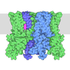
- EMDB-65347: Structure of hTRPV1 complexed with LIQ -

+
Open data


ID or keywords:

Loading...

-
Basic information

Entry
Database: EMDB / ID: EMD-65347
TitleStructure of hTRPV1 complexed with LIQ
Map data
Sample
  • Complex: TRPV1 complexed with LIQ
    • Protein or peptide: Transient receptor potential cation channel subfamily V member 1
  • Ligand: (2S)-3-(hexadecanoyloxy)-2-[(9Z)-octadec-9-enoyloxy]propyl 2-(trimethylammonio)ethyl phosphate
  • Ligand: Liquiritin
  • Ligand: 1-palmitoyl-2-oleoyl-sn-glycero-3-phosphoinositol
KeywordsComplex / Ion channel / MEMBRANE PROTEIN
Function / homology
Function and homology information


chemosensory behavior / response to capsazepine / sensory perception of mechanical stimulus / detection of temperature stimulus involved in thermoception / peptide secretion / temperature-gated ion channel activity / detection of chemical stimulus involved in sensory perception of pain / smooth muscle contraction involved in micturition / fever generation / thermoception ...chemosensory behavior / response to capsazepine / sensory perception of mechanical stimulus / detection of temperature stimulus involved in thermoception / peptide secretion / temperature-gated ion channel activity / detection of chemical stimulus involved in sensory perception of pain / smooth muscle contraction involved in micturition / fever generation / thermoception / excitatory extracellular ligand-gated monoatomic ion channel activity / dendritic spine membrane / diet induced thermogenesis / cellular response to alkaloid / TRP channels / intracellularly gated calcium channel activity / cellular response to ATP / detection of temperature stimulus involved in sensory perception of pain / behavioral response to pain / calcium ion import across plasma membrane / voltage-gated calcium channel activity / cellular response to acidic pH / extracellular ligand-gated monoatomic ion channel activity / phosphatidylinositol binding / lipid metabolic process / phosphoprotein binding / GABA-ergic synapse / calcium channel activity / calcium ion transmembrane transport / transmembrane signaling receptor activity / cellular response to heat / sensory perception of taste / protein homotetramerization / calmodulin binding / postsynaptic membrane / cell surface receptor signaling pathway / negative regulation of transcription by RNA polymerase II / ATP binding / membrane / metal ion binding / plasma membrane
Similarity search - Function
Transient receptor potential cation channel subfamily V member 1-4 / Transient receptor potential cation channel subfamily V / Ankyrin repeat / Ankyrin repeat profile. / Ankyrin repeats (3 copies) / Ankyrin repeat region circular profile. / ankyrin repeats / Ankyrin repeat / Ankyrin repeat-containing domain superfamily / Ion transport domain / Ion transport protein
Similarity search - Domain/homology
Transient receptor potential cation channel subfamily V member 1
Similarity search - Component
Biological speciesHomo sapiens (human)
Methodsingle particle reconstruction / cryo EM / Resolution: 3.26 Å
AuthorsMin YM / Zonglin DZ / Yang YY
Funding support China, 1 items
OrganizationGrant numberCountry
National Natural Science Foundation of China (NSFC) China
CitationJournal: Acta Crystallogr D Struct Biol / Year: 2019
Title: Macromolecular structure determination using X-rays, neutrons and electrons: recent developments in Phenix.
Authors: Dorothee Liebschner / Pavel V Afonine / Matthew L Baker / Gábor Bunkóczi / Vincent B Chen / Tristan I Croll / Bradley Hintze / Li Wei Hung / Swati Jain / Airlie J McCoy / Nigel W Moriarty ...Authors: Dorothee Liebschner / Pavel V Afonine / Matthew L Baker / Gábor Bunkóczi / Vincent B Chen / Tristan I Croll / Bradley Hintze / Li Wei Hung / Swati Jain / Airlie J McCoy / Nigel W Moriarty / Robert D Oeffner / Billy K Poon / Michael G Prisant / Randy J Read / Jane S Richardson / David C Richardson / Massimo D Sammito / Oleg V Sobolev / Duncan H Stockwell / Thomas C Terwilliger / Alexandre G Urzhumtsev / Lizbeth L Videau / Christopher J Williams / Paul D Adams /
Abstract: Diffraction (X-ray, neutron and electron) and electron cryo-microscopy are powerful methods to determine three-dimensional macromolecular structures, which are required to understand biological ...Diffraction (X-ray, neutron and electron) and electron cryo-microscopy are powerful methods to determine three-dimensional macromolecular structures, which are required to understand biological processes and to develop new therapeutics against diseases. The overall structure-solution workflow is similar for these techniques, but nuances exist because the properties of the reduced experimental data are different. Software tools for structure determination should therefore be tailored for each method. Phenix is a comprehensive software package for macromolecular structure determination that handles data from any of these techniques. Tasks performed with Phenix include data-quality assessment, map improvement, model building, the validation/rebuilding/refinement cycle and deposition. Each tool caters to the type of experimental data. The design of Phenix emphasizes the automation of procedures, where possible, to minimize repetitive and time-consuming manual tasks, while default parameters are chosen to encourage best practice. A graphical user interface provides access to many command-line features of Phenix and streamlines the transition between programs, project tracking and re-running of previous tasks.
History
DepositionJul 12, 2025-
Header (metadata) releaseJul 15, 2026-
Map releaseJul 15, 2026-
UpdateJul 15, 2026-
Current statusJul 15, 2026Processing site: PDBc / Status: Released

-
Structure visualization

Supplemental images

Downloads & links

-
Map

FileDownload / File: emd_65347.map.gz / Format: CCP4 / Size: 103 MB / Type: IMAGE STORED AS FLOATING POINT NUMBER (4 BYTES)
Projections & slices

Image control

Size
Brightness
Contrast
Others
AxesZ (Sec.)Y (Row.)X (Col.)
0.85 Å/pix.
x 300 pix.
= 255. Å
0.85 Å/pix.
x 300 pix.
= 255. Å
0.85 Å/pix.
x 300 pix.
= 255. Å

Surface

Projections

Slices (1/3)

Slices (1/2)

Slices (2/3)

Images are generated by Spider.

Voxel sizeX=Y=Z: 0.85 Å
Density
Contour LevelBy AUTHOR: 0.12
Minimum - Maximum-0.55262125 - 0.8542288
Average (Standard dev.)0.0015450333 (±0.028523091)
SymmetrySpace group: 1
Details

EMDB XML:

Map geometry
Axis orderXYZ
Origin000
Dimensions300300300
Spacing300300300
CellA=B=C: 255.0 Å
α=β=γ: 90.0 °

-
Supplemental data

-
Half map: #2

Fileemd_65347_half_map_1.map
Projections & Slices
AxesZYX

Projections

Slices (1/2)
Density Histograms

-
Half map: #1

Fileemd_65347_half_map_2.map
Projections & Slices
AxesZYX

Projections

Slices (1/2)
Density Histograms

-
Sample components

-
Entire : TRPV1 complexed with LIQ

EntireName: TRPV1 complexed with LIQ
Components
  • Complex: TRPV1 complexed with LIQ
    • Protein or peptide: Transient receptor potential cation channel subfamily V member 1
  • Ligand: (2S)-3-(hexadecanoyloxy)-2-[(9Z)-octadec-9-enoyloxy]propyl 2-(trimethylammonio)ethyl phosphate
  • Ligand: Liquiritin
  • Ligand: 1-palmitoyl-2-oleoyl-sn-glycero-3-phosphoinositol

-
Supramolecule #1: TRPV1 complexed with LIQ

SupramoleculeName: TRPV1 complexed with LIQ / type: complex / ID: 1 / Parent: 0 / Macromolecule list: #1
Source (natural)Organism: Homo sapiens (human)

-
Macromolecule #1: Transient receptor potential cation channel subfamily V member 1

MacromoleculeName: Transient receptor potential cation channel subfamily V member 1
type: protein_or_peptide / ID: 1 / Number of copies: 4 / Enantiomer: LEVO
Source (natural)Organism: Homo sapiens (human)
Molecular weightTheoretical: 95.063062 KDa
Recombinant expressionOrganism: Homo sapiens (human)
SequenceString: MKKWSSTDLG AAADPLQKDT CPDPLDGDPN SRPPPAKPQL STAKSRTRLF GKGDSEEAFP VDCPHEEGEL DSCPTITVSP VITIQRPGD GPTGARLLSQ DSVAASTEKT LRLYDRRSIF EAVAQNNCQD LESLLLFLQK SKKHLTDNEF KDPETGKTCL L KAMLNLHD ...String:
MKKWSSTDLG AAADPLQKDT CPDPLDGDPN SRPPPAKPQL STAKSRTRLF GKGDSEEAFP VDCPHEEGEL DSCPTITVSP VITIQRPGD GPTGARLLSQ DSVAASTEKT LRLYDRRSIF EAVAQNNCQD LESLLLFLQK SKKHLTDNEF KDPETGKTCL L KAMLNLHD GQNTTIPLLL EIARQTDSLK ELVNASYTDS YYKGQTALHI AIERRNMALV TLLVENGADV QAAAHGDFFK KT KGRPGFY FGELPLSLAA CTNQLGIVKF LLQNSWQTAD ISARDSVGNT VLHALVEVAD NTADNTKFVT SMYNEILMLG AKL HPTLKL EELTNKKGMT PLALAAGTGK IGVLAYILQR EIQEPECRHL SRKFTEWAYG PVHSSLYDLS CIDTCEKNSV LEVI AYSSS ETPNRHDMLL VEPLNRLLQD KWDRFVKRIF YFNFLVYCLY MIIFTMAAYY RPVDGLPPFK MEKTGDYFRV TGEIL SVLG GVYFFFRGIQ YFLQRRPSMK TLFVDSYSEM LFFLQSLFML ATVVLYFSHL KEYVASMVFS LALGWTNMLY YTRGFQ QMG IYAVMIEKMI LRDLCRFMFV YIVFLFGFST AVVTLIEDGK NDSLPSESTS HRWRGPACRP PDSSYNSLYS TCLELFK FT IGMGDLEFTE NYDFKAVFII LLLAYVILTY ILLLNMLIAL MGETVNKIAQ ESKNIWKLQR AITILDTEKS FLKCMRKA F RSGKLLQVGY TPDGKDDYRW CFRVDEVNWT TWNTNVGIIN EDPGNCEGVK RTLSFSLRSS RVSGRHWKNF ALVPLLREA SARDRQSAQP EEVYLRQFSG SLKPEDAEVF KSPAASGEK

UniProtKB: Transient receptor potential cation channel subfamily V member 1

-
Macromolecule #2: (2S)-3-(hexadecanoyloxy)-2-[(9Z)-octadec-9-enoyloxy]propyl 2-(tri...

MacromoleculeName: (2S)-3-(hexadecanoyloxy)-2-[(9Z)-octadec-9-enoyloxy]propyl 2-(trimethylammonio)ethyl phosphate
type: ligand / ID: 2 / Number of copies: 4 / Formula: POV
Molecular weightTheoretical: 760.076 Da
Chemical component information

ChemComp-POV:
(2S)-3-(hexadecanoyloxy)-2-[(9Z)-octadec-9-enoyloxy]propyl 2-(trimethylammonio)ethyl phosphate / phospholipid*YM

-
Macromolecule #3: Liquiritin

MacromoleculeName: Liquiritin / type: ligand / ID: 3 / Number of copies: 1 / Formula: A1ETR
Molecular weightTheoretical: 418.394 Da

-
Macromolecule #4: 1-palmitoyl-2-oleoyl-sn-glycero-3-phosphoinositol

MacromoleculeName: 1-palmitoyl-2-oleoyl-sn-glycero-3-phosphoinositol / type: ligand / ID: 4 / Number of copies: 3 / Formula: YBG
Molecular weightTheoretical: 837.069 Da
Chemical component information

ChemComp-YBG:
1-palmitoyl-2-oleoyl-sn-glycero-3-phosphoinositol

-
Experimental details

-
Structure determination

Methodcryo EM
Processingsingle particle reconstruction
Aggregation stateparticle

-
Sample preparation

BufferpH: 7.4
VitrificationCryogen name: ETHANE

-
Electron microscopy

MicroscopeTFS KRIOS
Image recordingFilm or detector model: FEI FALCON IV (4k x 4k) / Average electron dose: 50.0 e/Å2
Electron beamAcceleration voltage: 300 kV / Electron source: FIELD EMISSION GUN
Electron opticsIllumination mode: FLOOD BEAM / Imaging mode: BRIGHT FIELD / Nominal defocus max: 1.6 µm / Nominal defocus min: 1.0 µm
Experimental equipment
Model: Titan Krios / Image courtesy: FEI Company

+
Image processing

CTF correctionType: NONE
Startup modelType of model: PDB ENTRY
PDB model - PDB ID:
Final reconstructionResolution.type: BY AUTHOR / Resolution: 3.26 Å / Resolution method: FSC 0.143 CUT-OFF / Software - Name: cryoSPARC / Number images used: 36299
Initial angle assignmentType: MAXIMUM LIKELIHOOD
Final angle assignmentType: MAXIMUM LIKELIHOOD
FSC plot (resolution estimation)

+
About Yorodumi

-
News

-
Feb 9, 2022. New format data for meta-information of EMDB entries

New format data for meta-information of EMDB entries

  • Version 3 of the EMDB header file is now the official format.
  • The previous official version 1.9 will be removed from the archive.

Related info.:EMDB header

External links:wwPDB to switch to version 3 of the EMDB data model

-
Aug 12, 2020. Covid-19 info

Covid-19 info

URL: https://pdbj.org/emnavi/covid19.php

New page: Covid-19 featured information page in EM Navigator.

Related info.:Covid-19 info / Mar 5, 2020. Novel coronavirus structure data

+
Mar 5, 2020. Novel coronavirus structure data

Novel coronavirus structure data

Related info.:Yorodumi Speices / Aug 12, 2020. Covid-19 info

External links:COVID-19 featured content - PDBj / Molecule of the Month (242):Coronavirus Proteases

+
Jan 31, 2019. EMDB accession codes are about to change! (news from PDBe EMDB page)

EMDB accession codes are about to change! (news from PDBe EMDB page)

  • The allocation of 4 digits for EMDB accession codes will soon come to an end. Whilst these codes will remain in use, new EMDB accession codes will include an additional digit and will expand incrementally as the available range of codes is exhausted. The current 4-digit format prefixed with “EMD-” (i.e. EMD-XXXX) will advance to a 5-digit format (i.e. EMD-XXXXX), and so on. It is currently estimated that the 4-digit codes will be depleted around Spring 2019, at which point the 5-digit format will come into force.
  • The EM Navigator/Yorodumi systems omit the EMD- prefix.

Related info.:Q: What is EMD? / ID/Accession-code notation in Yorodumi/EM Navigator

External links:EMDB Accession Codes are Changing Soon! / Contact to PDBj

+
Jul 12, 2017. Major update of PDB

Major update of PDB

  • wwPDB released updated PDB data conforming to the new PDBx/mmCIF dictionary.
  • This is a major update changing the version number from 4 to 5, and with Remediation, in which all the entries are updated.
  • In this update, many items about electron microscopy experimental information are reorganized (e.g. em_software).
  • Now, EM Navigator and Yorodumi are based on the updated data.

External links:wwPDB Remediation / Enriched Model Files Conforming to OneDep Data Standards Now Available in the PDB FTP Archive

-
Yorodumi

Thousand views of thousand structures

  • Yorodumi is a browser for structure data from EMDB, PDB, SASBDB, etc.
  • This page is also the successor to EM Navigator detail page, and also detail information page/front-end page for Omokage search.
  • The word "yorodu" (or yorozu) is an old Japanese word meaning "ten thousand". "mi" (miru) is to see.

Related info.:EMDB / PDB / SASBDB / Comparison of 3 databanks / Yorodumi Search / Aug 31, 2016. New EM Navigator & Yorodumi / Yorodumi Papers / Jmol/JSmol / Function and homology information / Changes in new EM Navigator and Yorodumi

Read more