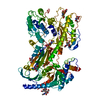
- EMDB-65160: Structure of human alpha-2/delta-1 with crisugabalin -
+
Open data
ID or keywords:
Loading...
-
Basic information
Entry
Database: EMDB / ID: EMD-65160
Title
Structure of human alpha-2/delta-1 with crisugabalin
Map data
Sample
Complex: Structure of human alpha-2/delta-1 with crisugabalin
Protein or peptide: Isoform 2 of Voltage-dependent calcium channel subunit alpha-2/delta-1
Ligand: 2-acetamido-2-deoxy-beta-D-glucopyranose
Ligand: Crisugabalin
Keywords
Complex / Alpha-2/delta-1 / Crisugabalin / MEMBRANE PROTEIN
Function / homology
Function and homology information
regulation of membrane repolarization during action potential / calcium ion transmembrane transport via high voltage-gated calcium channel / membrane depolarization during bundle of His cell action potential / L-type voltage-gated calcium channel complex / regulation of ventricular cardiac muscle cell membrane repolarization / cardiac muscle cell action potential involved in contraction / calcium ion transport into cytosol / regulation of calcium ion transmembrane transport via high voltage-gated calcium channel / voltage-gated calcium channel complex / Mechanical load activates signaling by PIEZO1 and integrins in osteocytes ...regulation of membrane repolarization during action potential / calcium ion transmembrane transport via high voltage-gated calcium channel / membrane depolarization during bundle of His cell action potential / L-type voltage-gated calcium channel complex / regulation of ventricular cardiac muscle cell membrane repolarization / cardiac muscle cell action potential involved in contraction / calcium ion transport into cytosol / regulation of calcium ion transmembrane transport via high voltage-gated calcium channel / voltage-gated calcium channel complex / Mechanical load activates signaling by PIEZO1 and integrins in osteocytes / regulation of calcium ion transport / regulation of heart rate by cardiac conduction / calcium ion import across plasma membrane / neuronal dense core vesicle / voltage-gated calcium channel activity / presynaptic active zone membrane / sarcoplasmic reticulum / GABA-ergic synapse / cellular response to amyloid-beta / calcium ion transport / extracellular exosome / metal ion binding / plasma membrane Similarity search - Function
VWA N-terminal / Voltage-dependent calcium channel, alpha-2/delta subunit, conserved region / VWA N-terminal / Neuronal voltage-dependent calcium channel alpha 2acd / : / von Willebrand factor type A domain / VWFA domain profile. / von Willebrand factor (vWF) type A domain / von Willebrand factor, type A / von Willebrand factor A-like domain superfamily Similarity search - Domain/homology
Journal: J Chem Inf Model / Year: 2026 Title: Structural and Computational Insights into the Mechanism of the Superior Pharmacological Activity of Crisugabalin: A Third-Generation Cavαδ1 Ligand. Authors: Zhaoqiang Chen / Xiaoli Gou / Qingyuan Meng / He Li / Yao Li / Zongjun Shi / Xinxin Li / Ju Wang / Abstract: Crisugabalin, a recently approved third-generation GABA analogue with a unique cage-like tricyclic scaffold, shows superior efficacy and safety over pregabalin and mirogabalin for treating ...Crisugabalin, a recently approved third-generation GABA analogue with a unique cage-like tricyclic scaffold, shows superior efficacy and safety over pregabalin and mirogabalin for treating neuropathic pain. Through integrated biophysical, structural, and computational approaches, we elucidate the molecular basis of its enhanced pharmacological profile. Dissociation kinetic studies revealed that crisugabalin exhibited the slowest dissociation kinetics from the αδ1 subunit (τ = 32.05, 80.00, 111.11 min for pregabalin, mirogabalin, and crisugabalin) but the fastest dissociation from the αδ2 subunit (τ = 8.70, 16.39, 5.78 min for pregabalin, mirogabalin, and crisugabalin). Cryo-EM structures demonstrated crisugabalin's superior binding affinity for αδ1 over gabapentin and l-leucine, driven by enhanced hydrogen bonding and hydrophobic contacts, alongside volumetric expansion of the l-leucine binding pocket. Molecular dynamics (MD) simulations identified significantly more persistent hydrogen bonding by crisugabalin (66.3% average occupancy) relative to pregabalin (28.3%). Random Acceleration Molecular Dynamics (RAMD) simulations revealed that ligand dissociation primarily proceeds via Pathway A (along the β2, β3, and β1 segments), and τRAMD calculations correctly ranked the ligand residence times, yielding values of 0.18 ns for pregabalin and 2.88 ns for crisugabalin. Furthermore, the binding free energies for pregabalin, mirogabalin, and crisugabalin were -21.64, -31.30, and -34.99 kcal/mol, calculated by MM/GBSA. The decomposition of the binding free energy components revealed that crisugabalin exhibits a dual-action mechanism characterized by enhanced hydrophobic interactions (-28.46 kcal/mol) and favorable entropic contributions (3.03 kcal/mol). This unique binding behavior stems from its cage-like tricyclic scaffold, an unprecedented substructure in drug molecules. These findings establish the cage-like tricyclic motif as a novel pharmacophore that simultaneously optimizes binding entropy and enthalpy, providing a blueprint for next-generation voltage-gated calcium channel modulators. MD, τRAMD, and MM-GBSA used in this study are powerful computational tools for rational drug design, particularly for optimizing compounds with prolonged target residence times.
In the structure databanks used in Yorodumi, some data are registered as the other names, "COVID-19 virus" and "2019-nCoV". Here are the details of the virus and the list of structure data.
Jan 31, 2019. EMDB accession codes are about to change! (news from PDBe EMDB page)
EMDB accession codes are about to change! (news from PDBe EMDB page)
The allocation of 4 digits for EMDB accession codes will soon come to an end. Whilst these codes will remain in use, new EMDB accession codes will include an additional digit and will expand incrementally as the available range of codes is exhausted. The current 4-digit format prefixed with “EMD-” (i.e. EMD-XXXX) will advance to a 5-digit format (i.e. EMD-XXXXX), and so on. It is currently estimated that the 4-digit codes will be depleted around Spring 2019, at which point the 5-digit format will come into force.
The EM Navigator/Yorodumi systems omit the EMD- prefix.
Related info.:Q: What is EMD? / ID/Accession-code notation in Yorodumi/EM Navigator
Yorodumi is a browser for structure data from EMDB, PDB, SASBDB, etc.
This page is also the successor to EM Navigator detail page, and also detail information page/front-end page for Omokage search.
The word "yorodu" (or yorozu) is an old Japanese word meaning "ten thousand". "mi" (miru) is to see.
Related info.:EMDB / PDB / SASBDB / Comparison of 3 databanks / Yorodumi Search / Aug 31, 2016. New EM Navigator & Yorodumi / Yorodumi Papers / Jmol/JSmol / Function and homology information / Changes in new EM Navigator and Yorodumi
 Movie
Movie Controller


 Open data
 Basic information
 Map data
 Sample
 Keywords
 Function and homology information
 Homo sapiens (human)
 Authors
 Citation
 Structure visualization
 Downloads & links
 emd_65160.png
 http://ftp.pdbj.org/pub/emdb/structures/EMD-65160


 Z (Sec.)
 Y (Row.)
 X (Col.)


 Sample components
 Processing
 Electron microscopy
 FIELD EMISSION GUN