 ムービー
ムービー コントローラー


+ データを開く

- 基本情報
基本情報
| 登録情報 | データベース: EMDB / ID: EMD-6512 | |||||||||
|---|---|---|---|---|---|---|---|---|---|---|
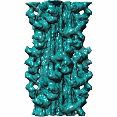
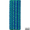
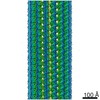
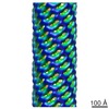
| タイトル | 3D reconstruction of tarantula thick filaments | |||||||||
 マップデータ マップデータ | 3D reconstruction of tarantula thick filament | |||||||||
 試料 試料 |
| |||||||||
 キーワード キーワード | single particle analysis / thick filament / muscle contraction | |||||||||
| 生物種 |  Grammostola rosea (クモ) Grammostola rosea (クモ) | |||||||||
| 手法 | 単粒子再構成法 / クライオ電子顕微鏡法 / 解像度: 13.0 Å | |||||||||
 データ登録者 データ登録者 | Yang S / Woodhead JL / Zhao F / Sulbaran G / Craig R | |||||||||
 引用 引用 | ジャーナル: J Struct Biol / 年: 2016 タイトル: An approach to improve the resolution of helical filaments with a large axial rise and flexible subunits. 著者: Shixin Yang / John L Woodhead / Fa-Qing Zhao / Guidenn Sulbarán / Roger Craig /  要旨: Single particle analysis is widely used for three-dimensional reconstruction of helical filaments. Near-atomic resolution has been obtained for several well-ordered filaments. However, it is still a ...Single particle analysis is widely used for three-dimensional reconstruction of helical filaments. Near-atomic resolution has been obtained for several well-ordered filaments. However, it is still a challenge to achieve high resolution for filaments with flexible subunits and a large axial rise per subunit relative to pixel size. Here, we describe an approach that improves the resolution in such cases. In filaments with a large axial rise, many segments must be shifted a long distance along the filament axis to match with a reference projection, potentially causing loss of alignment accuracy and hence resolution. In our study of myosin filaments, we overcame this problem by pre-determining the axial positions of myosin head crowns within segments to decrease the alignment error. In addition, homogeneous, well-ordered segments were selected from the raw data set by checking the assigned azimuthal rotation angle of segments in each filament against those expected for perfect helical symmetry. These procedures improved the resolution of the filament reconstruction from 30 Å to 13 Å. This approach could be useful in other helical filaments with a large axial rise and/or flexible subunits. | |||||||||
| 履歴 |
|
- 構造の表示
構造の表示
| ムービー |
 ムービービューア ムービービューア |
|---|---|
| 構造ビューア | EMマップ: SurfViewMolmilJmol/JSmol |
| 添付画像 |
- ダウンロードとリンク
-EMDBアーカイブ
| マップデータ | emd_6512.map.gz | 49.8 MB | EMDBマップデータ形式 | |
|---|---|---|---|---|
| ヘッダ (付随情報) | emd-6512-v30.xmlemd-6512.xml | 9.7 KB 9.7 KB | 表示 表示 | EMDBヘッダ |
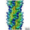
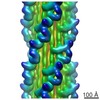
| 画像 |  400_6512.gif 400_6512.gif 80_6512.gif 80_6512.gif | 77.5 KB 5 KB | ||
| アーカイブディレクトリ |  http://ftp.pdbj.org/pub/emdb/structures/EMD-6512ftp://ftp.pdbj.org/pub/emdb/structures/EMD-6512 http://ftp.pdbj.org/pub/emdb/structures/EMD-6512ftp://ftp.pdbj.org/pub/emdb/structures/EMD-6512 | HTTPS FTP |
-関連構造データ
-リンク
| EMDBのページ | EMDB (EBI/PDBe) / EMDataResource |
|---|
-マップ
| ファイル | ダウンロード / ファイル: emd_6512.map.gz / 形式: CCP4 / 大きさ: 51.5 MB / タイプ: IMAGE STORED AS FLOATING POINT NUMBER (4 BYTES) | ||||||||||||||||||||||||||||||||||||||||||||||||||||||||||||||||||||
|---|---|---|---|---|---|---|---|---|---|---|---|---|---|---|---|---|---|---|---|---|---|---|---|---|---|---|---|---|---|---|---|---|---|---|---|---|---|---|---|---|---|---|---|---|---|---|---|---|---|---|---|---|---|---|---|---|---|---|---|---|---|---|---|---|---|---|---|---|---|
| 注釈 | 3D reconstruction of tarantula thick filament | ||||||||||||||||||||||||||||||||||||||||||||||||||||||||||||||||||||
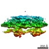
| 投影像・断面図 | 画像のコントロール
画像は Spider により作成 | ||||||||||||||||||||||||||||||||||||||||||||||||||||||||||||||||||||
| ボクセルのサイズ | X=Y=Z: 2.744 Å | ||||||||||||||||||||||||||||||||||||||||||||||||||||||||||||||||||||
| 密度 |
| ||||||||||||||||||||||||||||||||||||||||||||||||||||||||||||||||||||
| 対称性 | 空間群: 1 | ||||||||||||||||||||||||||||||||||||||||||||||||||||||||||||||||||||
| 詳細 | EMDB XML:
CCP4マップ ヘッダ情報:
| ||||||||||||||||||||||||||||||||||||||||||||||||||||||||||||||||||||
 Z (Sec.)
 Y (Row.)
 X (Col.)


-添付データ
- 試料の構成要素
-全体 : tarantula thick filament
| 全体 | 名称: tarantula thick filament |
|---|---|
| 要素 |
|
-超分子 #1000: tarantula thick filament
| 超分子 | 名称: tarantula thick filament / タイプ: sample / ID: 1000 / 集合状態: helical filaments of myosin II / Number unique components: 1 |
|---|---|
| 分子量 | 実験値: 520 KDa / 理論値: 520 KDa / 手法: Sedimentation |
-分子 #1: thick filament
| 分子 | 名称: thick filament / タイプ: protein_or_peptide / ID: 1 / Name.synonym: myosin filament / コピー数: 18 / 集合状態: helical filament / 組換発現: No / データベース: NCBI |
|---|---|
| 由来(天然) | 生物種: Grammostola rosea (クモ) / 別称: tarantula / 組織: leg / 細胞: striated muscle / Organelle: myofibril / 細胞中の位置: cytoskeleton |
| 分子量 | 実験値: 520 KDa / 理論値: 520 KDa |
-実験情報
-構造解析
| 手法 | クライオ電子顕微鏡法 |
|---|---|
 解析 解析 | 単粒子再構成法 |
| 試料の集合状態 | filament |
-試料調製
| 濃度 | 1.0 mg/mL |
|---|---|
| 緩衝液 | pH: 7 詳細: 100 mM NaCl, 3 mM MgCl2, 1 mM EGTA, 5 mM PIPES, 5 mM NaH2PO4, 1 mM NaN3 |
| グリッド | 詳細: Quantifoil (R2/2) 200 mesh grids, glow discharged |
| 凍結 | 凍結剤: ETHANE / チャンバー内湿度: 90 % / チャンバー内温度: 85 K / 装置: HOMEMADE PLUNGER / 手法: Blot for 2 seconds before plunging |
- 電子顕微鏡法
| 顕微鏡 | FEI TITAN KRIOS |
|---|---|
| 日付 | 2013年4月13日 |
| 撮影 | カテゴリ: CCD フィルム・検出器のモデル: GATAN ULTRASCAN 4000 (4k x 4k) 実像数: 690 / 平均電子線量: 20 e/Å2 |
| 電子線 | 加速電圧: 120 kV / 電子線源:  FIELD EMISSION GUN FIELD EMISSION GUN |
| 電子光学系 | 倍率(補正後): 109000 / 照射モード: FLOOD BEAM / 撮影モード: BRIGHT FIELD / Cs: 2.7 mm / 最大 デフォーカス(公称値): 4.5 µm / 最小 デフォーカス(公称値): 1.1 µm / 倍率(公称値): 59000 |
| 試料ステージ | 試料ホルダー: Nitrogen-cooled 試料ホルダーモデル: FEI TITAN KRIOS AUTOGRID HOLDER |
| 実験機器 |  モデル: Titan Krios / 画像提供: FEI Company |
-画像解析
| 詳細 | Cryo-EM images of tarantula thick filaments were processed using SPIDER and IHRSR |
|---|---|
| CTF補正 | 詳細: each particle |
| 最終 再構成 | 想定した対称性 - らせんパラメータ - Δz: 145 Å 想定した対称性 - らせんパラメータ - ΔΦ: 30 ° 想定した対称性 - らせんパラメータ - 軸対称性: C4 (4回回転対称) アルゴリズム: OTHER / 解像度のタイプ: BY AUTHOR / 解像度: 13.0 Å / 解像度の算出法: OTHER / ソフトウェア - 名称: SPIDER, IHRSR / 使用した粒子像数: 3174 |
