 Movie
Movie Controller


+ Open data

- Basic information
| Entry | Database: EMDB / ID: EMD-6514 | |||||||||
|---|---|---|---|---|---|---|---|---|---|---|
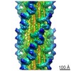
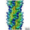
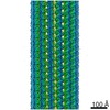
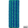
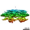
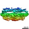
| Title | 3D reconstruction of tarantula thick filaments | |||||||||
 Map data Map data | 3D reconstruction of Tarantula thick filament at 30 Angstrom resolution | |||||||||
 Sample Sample |
| |||||||||
 Keywords Keywords | single particle analysis / thick filament / muscle contraction | |||||||||
| Biological species |  Grammostola rosea (Chilean rose tarantula) Grammostola rosea (Chilean rose tarantula) | |||||||||
| Method | single particle reconstruction / cryo EM / Resolution: 30.0 Å | |||||||||
 Authors Authors | Yang S / Woodhead JL / Zhao F / Sulbaran G / Craig R | |||||||||
 Citation Citation | Journal: J Struct Biol / Year: 2016 Title: An approach to improve the resolution of helical filaments with a large axial rise and flexible subunits. Authors: Shixin Yang / John L Woodhead / Fa-Qing Zhao / Guidenn Sulbarán / Roger Craig /  Abstract: Single particle analysis is widely used for three-dimensional reconstruction of helical filaments. Near-atomic resolution has been obtained for several well-ordered filaments. However, it is still a ...Single particle analysis is widely used for three-dimensional reconstruction of helical filaments. Near-atomic resolution has been obtained for several well-ordered filaments. However, it is still a challenge to achieve high resolution for filaments with flexible subunits and a large axial rise per subunit relative to pixel size. Here, we describe an approach that improves the resolution in such cases. In filaments with a large axial rise, many segments must be shifted a long distance along the filament axis to match with a reference projection, potentially causing loss of alignment accuracy and hence resolution. In our study of myosin filaments, we overcame this problem by pre-determining the axial positions of myosin head crowns within segments to decrease the alignment error. In addition, homogeneous, well-ordered segments were selected from the raw data set by checking the assigned azimuthal rotation angle of segments in each filament against those expected for perfect helical symmetry. These procedures improved the resolution of the filament reconstruction from 30 Å to 13 Å. This approach could be useful in other helical filaments with a large axial rise and/or flexible subunits. | |||||||||
| History |
|
- Structure visualization
| Movie |
 Movie viewer Movie viewer |
|---|---|
| Structure viewer | EM map: SurfViewMolmilJmol/JSmol |
| Supplemental images |
- Downloads & links
-EMDB archive
| Map data | emd_6514.map.gz | 49.5 MB | EMDB map data format | |
|---|---|---|---|---|
| Header (meta data) | emd-6514-v30.xmlemd-6514.xml | 9.8 KB 9.8 KB | Display Display | EMDB header |
| Images |  400_6514.gif 400_6514.gif 80_6514.gif 80_6514.gif | 75.5 KB 5.2 KB | ||
| Archive directory |  http://ftp.pdbj.org/pub/emdb/structures/EMD-6514ftp://ftp.pdbj.org/pub/emdb/structures/EMD-6514 http://ftp.pdbj.org/pub/emdb/structures/EMD-6514ftp://ftp.pdbj.org/pub/emdb/structures/EMD-6514 | HTTPS FTP |
-Related structure data
-Links
| EMDB pages | EMDB (EBI/PDBe) / EMDataResource |
|---|
-Map
| File | Download / File: emd_6514.map.gz / Format: CCP4 / Size: 51.5 MB / Type: IMAGE STORED AS FLOATING POINT NUMBER (4 BYTES) | ||||||||||||||||||||||||||||||||||||||||||||||||||||||||||||||||||||
|---|---|---|---|---|---|---|---|---|---|---|---|---|---|---|---|---|---|---|---|---|---|---|---|---|---|---|---|---|---|---|---|---|---|---|---|---|---|---|---|---|---|---|---|---|---|---|---|---|---|---|---|---|---|---|---|---|---|---|---|---|---|---|---|---|---|---|---|---|---|
| Annotation | 3D reconstruction of Tarantula thick filament at 30 Angstrom resolution | ||||||||||||||||||||||||||||||||||||||||||||||||||||||||||||||||||||
| Projections & slices | Image control
Images are generated by Spider. | ||||||||||||||||||||||||||||||||||||||||||||||||||||||||||||||||||||
| Voxel size | X=Y=Z: 2.744 Å | ||||||||||||||||||||||||||||||||||||||||||||||||||||||||||||||||||||
| Density |
| ||||||||||||||||||||||||||||||||||||||||||||||||||||||||||||||||||||
| Symmetry | Space group: 1 | ||||||||||||||||||||||||||||||||||||||||||||||||||||||||||||||||||||
| Details | EMDB XML:
CCP4 map header:
| ||||||||||||||||||||||||||||||||||||||||||||||||||||||||||||||||||||
 Z (Sec.)
 Y (Row.)
 X (Col.)


-Supplemental data
- Sample components
-Entire : tarantula thick filament
| Entire | Name: tarantula thick filament |
|---|---|
| Components |
|
-Supramolecule #1000: tarantula thick filament
| Supramolecule | Name: tarantula thick filament / type: sample / ID: 1000 / Oligomeric state: helical filaments of myosin II / Number unique components: 1 |
|---|---|
| Molecular weight | Experimental: 520 KDa / Theoretical: 520 KDa / Method: Sedimentation |
-Macromolecule #1: thick filament
| Macromolecule | Name: thick filament / type: protein_or_peptide / ID: 1 / Name.synonym: myosin filament / Number of copies: 18 / Oligomeric state: helical filament / Recombinant expression: No / Database: NCBI |
|---|---|
| Source (natural) | Organism: Grammostola rosea (Chilean rose tarantula) / synonym: tarantula / Tissue: leg / Cell: striated muscle / Organelle: myofibril / Location in cell: cytoskeleton |
| Molecular weight | Experimental: 520 KDa / Theoretical: 520 KDa |
-Experimental details
-Structure determination
| Method | cryo EM |
|---|---|
 Processing Processing | single particle reconstruction |
| Aggregation state | filament |
-Sample preparation
| Concentration | 1.0 mg/mL |
|---|---|
| Buffer | pH: 7 Details: 100 mM NaCl, 3 mM MgCl2, 1 mM EGTA, 5 mM PIPES, 5 mM NaH2PO4, 1 mM NaN3 |
| Grid | Details: Quantifoil (R2/2) 200 mesh grids, glow discharged |
| Vitrification | Cryogen name: ETHANE / Chamber humidity: 90 % / Chamber temperature: 85 K / Instrument: HOMEMADE PLUNGER / Method: Blot for 2 seconds before plunging |
- Electron microscopy
| Microscope | FEI TITAN KRIOS |
|---|---|
| Date | Apr 13, 2013 |
| Image recording | Category: CCD / Film or detector model: GATAN ULTRASCAN 4000 (4k x 4k) / Number real images: 690 / Average electron dose: 20 e/Å2 |
| Electron beam | Acceleration voltage: 120 kV / Electron source:  FIELD EMISSION GUN FIELD EMISSION GUN |
| Electron optics | Calibrated magnification: 109000 / Illumination mode: FLOOD BEAM / Imaging mode: BRIGHT FIELD / Cs: 2.7 mm / Nominal defocus max: 4.5 µm / Nominal defocus min: 1.1 µm / Nominal magnification: 59000 |
| Sample stage | Specimen holder: Nitrogen-cooled / Specimen holder model: FEI TITAN KRIOS AUTOGRID HOLDER |
| Experimental equipment |  Model: Titan Krios / Image courtesy: FEI Company |
-Image processing
| Details | Cryo-EM images of tarantula thick filaments were processed using SPIDER and IHRSR |
|---|---|
| CTF correction | Details: each particle |
| Final reconstruction | Applied symmetry - Helical parameters - Δz: 145 Å Applied symmetry - Helical parameters - Δ&Phi: 30 ° Applied symmetry - Helical parameters - Axial symmetry: C4 (4 fold cyclic) Algorithm: OTHER / Resolution.type: BY AUTHOR / Resolution: 30.0 Å / Resolution method: OTHER / Software - Name: SPIDER, IHRSR Details: Particles were not pre-centered. All particles were used for reconstruction. Number images used: 3174 |
