+ データを開く
- 基本情報
基本情報
| 登録情報 | データベース: EMDB / ID: EMD-6510 | |||||||||
|---|---|---|---|---|---|---|---|---|---|---|
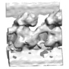
| タイトル | Docking complex-independent alignment of outer dynein arms with 24-nm periodicity | |||||||||
 マップデータ マップデータ | Streptavidin-labeled DC2-M412BCCP axoneme | |||||||||
 試料 試料 |
| |||||||||
 キーワード キーワード | cilia and flagella / axoneme / outer dynein arm / docking complex | |||||||||
| 生物種 |  | |||||||||
| 手法 | サブトモグラム平均法 / クライオ電子顕微鏡法 / 解像度: 47.0 Å | |||||||||
 データ登録者 データ登録者 | Oda T / Abe T / Yanagisawa H / Kikkawa M | |||||||||
 引用 引用 |  ジャーナル: J Cell Sci / 年: 2016 ジャーナル: J Cell Sci / 年: 2016タイトル: Docking-complex-independent alignment of Chlamydomonas outer dynein arms with 24-nm periodicity in vitro. 著者: Toshiyuki Oda / Tatsuki Abe / Haruaki Yanagisawa / Masahide Kikkawa /  要旨: The docking complex is a molecular complex necessary for assembly of outer dynein arms (ODAs) on the axonemal doublet microtubules (DMTs) in cilia and flagella. The docking complex is hypothesized to ...The docking complex is a molecular complex necessary for assembly of outer dynein arms (ODAs) on the axonemal doublet microtubules (DMTs) in cilia and flagella. The docking complex is hypothesized to be a 24-nm molecular ruler because ODAs align along the DMTs with 24-nm periodicity. In this study, we rigorously tested this hypothesis using structural and genetic methods. We found that the ODAs can bind to DMTs and porcine microtubules with 24-nm periodicities even in the absence of the docking complexin vitro Using cryo-electron tomography and structural labeling, we observed that the docking complex took an unexpectedly flexible conformation and did not lie along the length of DMTs. In the absence of docking complex, ODAs were released from the DMT at relatively low ionic strength conditions, suggesting that the docking complex strengthens the electrostatic interactions between the ODA and DMT. Based on these results, we conclude that the docking complex serves as a flexible stabilizer of the ODA rather than as a molecular ruler. | |||||||||
| 履歴 |
|
- 構造の表示
構造の表示
| ムービー |
 ムービービューア ムービービューア |
|---|---|
| 構造ビューア | EMマップ:  SurfView SurfView Molmil Molmil Jmol/JSmol Jmol/JSmol |
| 添付画像 |
- ダウンロードとリンク
-EMDBアーカイブ
| マップデータ |  emd_6510.map.gz emd_6510.map.gz | 985.1 KB |  EMDBマップデータ形式 EMDBマップデータ形式 | |
|---|---|---|---|---|
| ヘッダ (付随情報) |  emd-6510-v30.xml emd-6510-v30.xml emd-6510.xml emd-6510.xml | 8.6 KB 8.6 KB | 表示 表示 |  EMDBヘッダ EMDBヘッダ |
| 画像 |  400_6510.gif 400_6510.gif 80_6510.gif 80_6510.gif | 53.5 KB 4.5 KB | ||
| アーカイブディレクトリ |  http://ftp.pdbj.org/pub/emdb/structures/EMD-6510 http://ftp.pdbj.org/pub/emdb/structures/EMD-6510 ftp://ftp.pdbj.org/pub/emdb/structures/EMD-6510 ftp://ftp.pdbj.org/pub/emdb/structures/EMD-6510 | HTTPS FTP |
-検証レポート
| 文書・要旨 |  emd_6510_validation.pdf.gz emd_6510_validation.pdf.gz | 77.9 KB | 表示 |  EMDB検証レポート EMDB検証レポート |
|---|---|---|---|---|
| 文書・詳細版 |  emd_6510_full_validation.pdf.gz emd_6510_full_validation.pdf.gz | 77 KB | 表示 | |
| XML形式データ |  emd_6510_validation.xml.gz emd_6510_validation.xml.gz | 494 B | 表示 | |
| アーカイブディレクトリ |  https://ftp.pdbj.org/pub/emdb/validation_reports/EMD-6510 https://ftp.pdbj.org/pub/emdb/validation_reports/EMD-6510 ftp://ftp.pdbj.org/pub/emdb/validation_reports/EMD-6510 ftp://ftp.pdbj.org/pub/emdb/validation_reports/EMD-6510 | HTTPS FTP |
-関連構造データ
- リンク
リンク
| EMDBのページ |  EMDB (EBI/PDBe) / EMDB (EBI/PDBe) /  EMDataResource EMDataResource |
|---|
- マップ
マップ
| ファイル |  ダウンロード / ファイル: emd_6510.map.gz / 形式: CCP4 / 大きさ: 1.3 MB / タイプ: IMAGE STORED AS FLOATING POINT NUMBER (4 BYTES) ダウンロード / ファイル: emd_6510.map.gz / 形式: CCP4 / 大きさ: 1.3 MB / タイプ: IMAGE STORED AS FLOATING POINT NUMBER (4 BYTES) | ||||||||||||||||||||||||||||||||||||||||||||||||||||||||||||||||||||
|---|---|---|---|---|---|---|---|---|---|---|---|---|---|---|---|---|---|---|---|---|---|---|---|---|---|---|---|---|---|---|---|---|---|---|---|---|---|---|---|---|---|---|---|---|---|---|---|---|---|---|---|---|---|---|---|---|---|---|---|---|---|---|---|---|---|---|---|---|---|
| 注釈 | Streptavidin-labeled DC2-M412BCCP axoneme | ||||||||||||||||||||||||||||||||||||||||||||||||||||||||||||||||||||
| 投影像・断面図 | 画像のコントロール
画像は Spider により作成 | ||||||||||||||||||||||||||||||||||||||||||||||||||||||||||||||||||||
| ボクセルのサイズ | X=Y=Z: 6.07 Å | ||||||||||||||||||||||||||||||||||||||||||||||||||||||||||||||||||||
| 密度 |
| ||||||||||||||||||||||||||||||||||||||||||||||||||||||||||||||||||||
| 対称性 | 空間群: 1 | ||||||||||||||||||||||||||||||||||||||||||||||||||||||||||||||||||||
| 詳細 | EMDB XML:
CCP4マップ ヘッダ情報:
| ||||||||||||||||||||||||||||||||||||||||||||||||||||||||||||||||||||
-添付データ
- 試料の構成要素
-全体 : Streptavidin-labeled ODA structure from DC2-M412BCCP axoneme
| 全体 | 名称: Streptavidin-labeled ODA structure from DC2-M412BCCP axoneme |
|---|---|
| 要素 |
|
-超分子 #1000: Streptavidin-labeled ODA structure from DC2-M412BCCP axoneme
| 超分子 | 名称: Streptavidin-labeled ODA structure from DC2-M412BCCP axoneme タイプ: sample / ID: 1000 / Number unique components: 1 |
|---|
-超分子 #1: axoneme
| 超分子 | 名称: axoneme / タイプ: organelle_or_cellular_component / ID: 1 / 組換発現: No / データベース: NCBI |
|---|---|
| 由来(天然) | 生物種:  Organelle: flagella |
-実験情報
-構造解析
| 手法 | クライオ電子顕微鏡法 |
|---|---|
 解析 解析 | サブトモグラム平均法 |
| 試料の集合状態 | cell |
- 試料調製
試料調製
| 濃度 | 0.01 mg/mL |
|---|---|
| 緩衝液 | pH: 7.2 詳細: 30 mM Hepes-NaOH pH 7.2, 5 mM MgCl2, 1 mM dithiothreitol, 1 mM EGTA, 50 mM K-acetate |
| グリッド | 詳細: 300 mesh copper grid, holey carbon |
| 凍結 | 凍結剤: ETHANE / チャンバー内湿度: 90 % / チャンバー内温度: 93 K / 装置: LEICA EM GP / 手法: Blot for 5 seconds before plunging |
- 電子顕微鏡法
| 顕微鏡 | OTHER |
|---|---|
| 特殊光学系 | エネルギーフィルター - 名称: Omega エネルギーフィルター - エネルギー下限: 0.0 eV エネルギーフィルター - エネルギー上限: 20.0 eV |
| 日付 | 2015年2月20日 |
| 撮影 | カテゴリ: CCD フィルム・検出器のモデル: TVIPS TEMCAM-F416 (4k x 4k) 平均電子線量: 100 e/Å2 |
| 電子線 | 加速電圧: 300 kV / 電子線源:  FIELD EMISSION GUN FIELD EMISSION GUN |
| 電子光学系 | 照射モード: FLOOD BEAM / 撮影モード: BRIGHT FIELD / Cs: 2.2 mm / 最大 デフォーカス(公称値): 9.0 µm / 最小 デフォーカス(公称値): 6.0 µm / 倍率(公称値): 25700 |
| 試料ステージ | 試料ホルダーモデル: GATAN LIQUID NITROGEN / Tilt series - Axis1 - Min angle: -60 ° / Tilt series - Axis1 - Max angle: 60 ° |
- 画像解析
画像解析
| 詳細 | Number of tilts (projections) used in 3D reconstruction: 60 Tomographic tilt angle increment: 2 |
|---|---|
| 最終 再構成 | 解像度のタイプ: BY AUTHOR / 解像度: 47.0 Å / 解像度の算出法: OTHER / ソフトウェア - 名称: IMOD, PEET / 使用したサブトモグラム数: 936 |
 ムービー
ムービー コントローラー


 Z (Sec.)
 Y (Row.)
 X (Col.)


