Journal: Structure / Year: 2016 Title: Ring Separation Highlights the Protein-Folding Mechanism Used by the Phage EL-Encoded Chaperonin. Authors: Sudheer K Molugu / Zacariah L Hildenbrand / David Gene Morgan / Michael B Sherman / Lilin He / Costa Georgopoulos / Natalia V Sernova / Lidia P Kurochkina / Vadim V Mesyanzhinov / Konstantin ...Authors: Sudheer K Molugu / Zacariah L Hildenbrand / David Gene Morgan / Michael B Sherman / Lilin He / Costa Georgopoulos / Natalia V Sernova / Lidia P Kurochkina / Vadim V Mesyanzhinov / Konstantin A Miroshnikov / Ricardo A Bernal / Abstract: Chaperonins are ubiquitous, ATP-dependent protein-folding molecular machines that are essential for all forms of life. Bacteriophage φEL encodes its own chaperonin to presumably fold exceedingly ...Chaperonins are ubiquitous, ATP-dependent protein-folding molecular machines that are essential for all forms of life. Bacteriophage φEL encodes its own chaperonin to presumably fold exceedingly large viral proteins via profoundly different nucleotide-binding conformations. Our structural investigations indicate that ATP likely binds to both rings simultaneously and that a misfolded substrate acts as the trigger for ATP hydrolysis. More importantly, the φEL complex dissociates into two single rings resulting from an evolutionarily altered residue in the highly conserved ATP-binding pocket. Conformational changes also more than double the volume of the single-ring internal chamber such that larger viral proteins are accommodated. This is illustrated by the fact that φEL is capable of folding β-galactosidase, a 116-kDa protein. Collectively, the architecture and protein-folding mechanism of the φEL chaperonin are significantly different from those observed in group I and II chaperonins.
History
Deposition
Oct 21, 2015
-
Header (metadata) release
Mar 30, 2016
-
Map release
Mar 30, 2016
-
Update
Apr 27, 2016
-
Current status
Apr 27, 2016
Processing site: RCSB / Status: Released
-
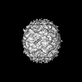
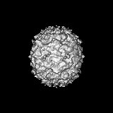
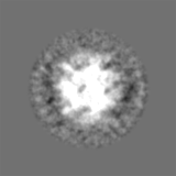
Structure visualization
Movie
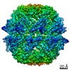
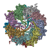
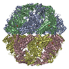
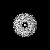
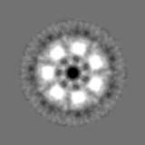
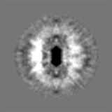
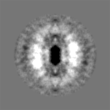
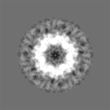
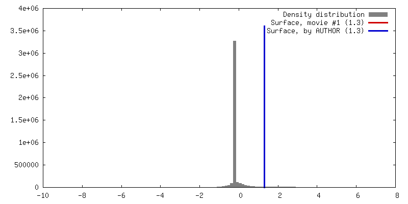
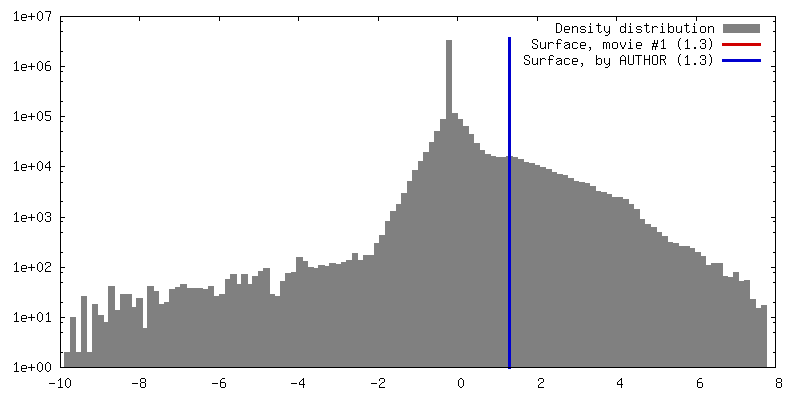
Surface view with section colored by density value
Organism: Pseudomonas phage EL (virus) / synonym: Bacteriophage EL
Molecular weight
Theoretical: 61.6811 KDa
Recombinant expression
Organism: Escherichia coli (E. coli) / Recombinant plasmid: pET22
Sequence
UniProtKB: Putative GroEL-like chaperonine protein
-
Experimental details
-
Structure determination
Method
cryo EM
Processing
single particle reconstruction
Aggregation state
particle
-
Sample preparation
Concentration
2.5 mg/mL
Buffer
pH: 7.5 Details: 50 mM HEPES, pH 7.5, 150 mM NaCl, 2 mM ADP, 2 mM EDTA
Grid
Details: Quantifoil R2/2 grids glow-discharged in air for 1 minute
Vitrification
Cryogen name: ETHANE / Chamber humidity: 80 % / Chamber temperature: 100 K / Instrument: HOMEMADE PLUNGER / Method: Blot for 2-3 seconds before plunging
-
Electron microscopy
Microscope
JEOL 2200FS
Temperature
Min: 88 K / Max: 103 K / Average: 100 K
Date
Aug 1, 2010
Image recording
Category: CCD / Film or detector model: GATAN ULTRASCAN 4000 (4k x 4k) / Digitization - Sampling interval: 12 µm / Number real images: 100 / Average electron dose: 10 e/Å2 / Bits/pixel: 8
Electron beam
Acceleration voltage: 200 kV / Electron source: FIELD EMISSION GUN
In the structure databanks used in Yorodumi, some data are registered as the other names, "COVID-19 virus" and "2019-nCoV". Here are the details of the virus and the list of structure data.
Jan 31, 2019. EMDB accession codes are about to change! (news from PDBe EMDB page)
EMDB accession codes are about to change! (news from PDBe EMDB page)
The allocation of 4 digits for EMDB accession codes will soon come to an end. Whilst these codes will remain in use, new EMDB accession codes will include an additional digit and will expand incrementally as the available range of codes is exhausted. The current 4-digit format prefixed with “EMD-” (i.e. EMD-XXXX) will advance to a 5-digit format (i.e. EMD-XXXXX), and so on. It is currently estimated that the 4-digit codes will be depleted around Spring 2019, at which point the 5-digit format will come into force.
The EM Navigator/Yorodumi systems omit the EMD- prefix.
Related info.:Q: What is EMD? / ID/Accession-code notation in Yorodumi/EM Navigator
Yorodumi is a browser for structure data from EMDB, PDB, SASBDB, etc.
This page is also the successor to EM Navigator detail page, and also detail information page/front-end page for Omokage search.
The word "yorodu" (or yorozu) is an old Japanese word meaning "ten thousand". "mi" (miru) is to see.
Related info.:EMDB / PDB / SASBDB / Comparison of 3 databanks / Yorodumi Search / Aug 31, 2016. New EM Navigator & Yorodumi / Yorodumi Papers / Jmol/JSmol / Function and homology information / Changes in new EM Navigator and Yorodumi
 Movie
Movie Controller

 Yorodumi
 Open data
 Basic information
 Map data
 Sample
 Keywords
 Function and homology information
 Pseudomonas phage EL (virus)
 Authors
 Citation

 Structure visualization
 Downloads & links
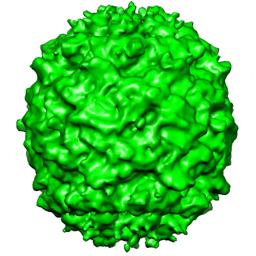 emd_6494.jpg
 http://ftp.pdbj.org/pub/emdb/structures/EMD-6494


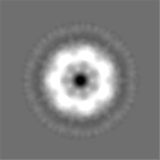
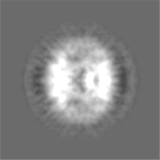
 Z (Sec.)
 Y (Row.)
 X (Col.)


 Sample components
 Processing
 Electron microscopy
 FIELD EMISSION GUN