[English] 日本語
 Yorodumi
- EMDB-64825: Structure of FtsZ1 in complex with GMPCPP from Candidatus Odinarc... -
+ Open data
- Basic information
| Entry |  | |||||||||
|---|---|---|---|---|---|---|---|---|---|---|
| Title | Structure of FtsZ1 in complex with GMPCPP from Candidatus Odinarchaeota | |||||||||
 Map data Map data | This is the final sharpened map with helical symmetry imposed. The contour level is based on values shown in chimera | |||||||||
 Sample Sample |
| |||||||||
 Keywords Keywords | Cell division Protein / CELL CYCLE | |||||||||
| Function / homology |  Function and homology information Function and homology informationFtsZ-dependent cytokinesis / cell division site / protein polymerization / GTPase activity / GTP binding / cytoplasm Similarity search - Function | |||||||||
| Biological species |  Candidatus Odinarchaeota (archaea) Candidatus Odinarchaeota (archaea) | |||||||||
| Method | helical reconstruction / cryo EM / Resolution: 3.6 Å | |||||||||
 Authors Authors | Bose S / Kutti RV | |||||||||
| Funding support |  India, 1 items India, 1 items
| |||||||||
 Citation Citation |  Journal: EMBO J / Year: 2025 Journal: EMBO J / Year: 2025Title: Distinct filament morphology and membrane tethering features of the dual FtsZ paralogs in Odinarchaeota. Authors: Jayanti Kumari / Akhilesh Uthaman / Sucharita Bose / Ananya Kundu / Vaibhav Sharma / Soumyajit Dutta / Anubhav Dhar / Srijita Roy / Ramanujam Srinivasan / Samay Pande / Kutti R Vinothkumar / ...Authors: Jayanti Kumari / Akhilesh Uthaman / Sucharita Bose / Ananya Kundu / Vaibhav Sharma / Soumyajit Dutta / Anubhav Dhar / Srijita Roy / Ramanujam Srinivasan / Samay Pande / Kutti R Vinothkumar / Pananghat Gayathri / Saravanan Palani /  Abstract: The Asgard phylum has emerged as a model to study eukaryogenesis because of their close relatedness with the eukaryotes. In this study, we use FtsZ proteins from a member of the class Odinarchaeia as ...The Asgard phylum has emerged as a model to study eukaryogenesis because of their close relatedness with the eukaryotes. In this study, we use FtsZ proteins from a member of the class Odinarchaeia as representatives to investigate the probable origin, evolution, and assembly of the FtsZ/tubulin protein superfamily in Asgard archaea. We performed a comparative analysis of the biochemical properties and cytoskeletal assembly of FtsZ1 and FtsZ2, the two FtsZ isoforms in the Odinarchaeota metagenome. Our electron microscopy analysis reveals that OdinFtsZ1 assembles into curved single protofilaments, while OdinFtsZ2 forms stacked spiral ring-like structures. Upon sequence analysis, we identified an N-terminal amphipathic helix in OdinFtsZ1, which mediates direct membrane tethering. In contrast, OdinFtsZ2 is recruited to the membrane by the anchor OdinSepF via OdinFtsZ2's C-terminal tail. Overall, we report the presence of two distant evolutionary paralogs of FtsZ in Odinarchaeota, with distinct filament assemblies and differing modes of membrane targeting. Our findings highlight the diversity of FtsZ proteins in the archaeal phylum Asgardarchaeota, providing valuable insights into the evolution and differentiation of tubulin-family proteins. | |||||||||
| History |
|
- Structure visualization
| Supplemental images |
|---|
- Downloads & links
-EMDB archive
| Map data |  emd_64825.map.gz emd_64825.map.gz | 8 MB |  EMDB map data format EMDB map data format | |
|---|---|---|---|---|
| Header (meta data) |  emd-64825-v30.xml emd-64825-v30.xml emd-64825.xml emd-64825.xml | 21.6 KB 21.6 KB | Display Display |  EMDB header EMDB header |
| FSC (resolution estimation) |  emd_64825_fsc.xml emd_64825_fsc.xml | 8.4 KB | Display |  FSC data file FSC data file |
| Images |  emd_64825.png emd_64825.png | 10 KB | ||
| Filedesc metadata |  emd-64825.cif.gz emd-64825.cif.gz | 6.8 KB | ||
| Others |  emd_64825_half_map_1.map.gz emd_64825_half_map_1.map.gz emd_64825_half_map_2.map.gz emd_64825_half_map_2.map.gz | 59.5 MB 59.5 MB | ||
| Archive directory |  http://ftp.pdbj.org/pub/emdb/structures/EMD-64825 http://ftp.pdbj.org/pub/emdb/structures/EMD-64825 ftp://ftp.pdbj.org/pub/emdb/structures/EMD-64825 ftp://ftp.pdbj.org/pub/emdb/structures/EMD-64825 | HTTPS FTP |
-Related structure data
| Related structure data |  9v7vMC M: atomic model generated by this map C: citing same article ( |
|---|---|
| Similar structure data | Similarity search - Function & homology  F&H Search F&H Search |
- Links
Links
| EMDB pages |  EMDB (EBI/PDBe) / EMDB (EBI/PDBe) /  EMDataResource EMDataResource |
|---|---|
| Related items in Molecule of the Month |
- Map
Map
| File |  Download / File: emd_64825.map.gz / Format: CCP4 / Size: 64 MB / Type: IMAGE STORED AS FLOATING POINT NUMBER (4 BYTES) Download / File: emd_64825.map.gz / Format: CCP4 / Size: 64 MB / Type: IMAGE STORED AS FLOATING POINT NUMBER (4 BYTES) | ||||||||||||||||||||||||||||||||||||
|---|---|---|---|---|---|---|---|---|---|---|---|---|---|---|---|---|---|---|---|---|---|---|---|---|---|---|---|---|---|---|---|---|---|---|---|---|---|
| Annotation | This is the final sharpened map with helical symmetry imposed. The contour level is based on values shown in chimera | ||||||||||||||||||||||||||||||||||||
| Projections & slices | Image control
Images are generated by Spider. | ||||||||||||||||||||||||||||||||||||
| Voxel size | X=Y=Z: 1.07 Å | ||||||||||||||||||||||||||||||||||||
| Density |
| ||||||||||||||||||||||||||||||||||||
| Symmetry | Space group: 1 | ||||||||||||||||||||||||||||||||||||
| Details | EMDB XML:
|
-Supplemental data
-Half map: This is half map A. This is unsharpened map....
| File | emd_64825_half_map_1.map | ||||||||||||
|---|---|---|---|---|---|---|---|---|---|---|---|---|---|
| Annotation | This is half map_A. This is unsharpened map. The contour level is based on values shown in chimera | ||||||||||||
| Projections & Slices |
| ||||||||||||
| Density Histograms |
-Half map: This is half map B. This is unsharpened map....
| File | emd_64825_half_map_2.map | ||||||||||||
|---|---|---|---|---|---|---|---|---|---|---|---|---|---|
| Annotation | This is half map_B. This is unsharpened map. The contour level is based on values shown in chimera | ||||||||||||
| Projections & Slices |
| ||||||||||||
| Density Histograms |
- Sample components
-Entire : FtsZ1 protein in complex with GMPCPP
| Entire | Name: FtsZ1 protein in complex with GMPCPP |
|---|---|
| Components |
|
-Supramolecule #1: FtsZ1 protein in complex with GMPCPP
| Supramolecule | Name: FtsZ1 protein in complex with GMPCPP / type: complex / ID: 1 / Parent: 0 / Macromolecule list: #1 |
|---|---|
| Source (natural) | Organism:  Candidatus Odinarchaeota (archaea) Candidatus Odinarchaeota (archaea) |
| Molecular weight | Theoretical: 8.8 kDa/nm |
-Macromolecule #1: Cell division protein FtsZ
| Macromolecule | Name: Cell division protein FtsZ / type: protein_or_peptide / ID: 1 / Number of copies: 4 / Enantiomer: LEVO |
|---|---|
| Source (natural) | Organism:  Candidatus Odinarchaeota (archaea) Candidatus Odinarchaeota (archaea) |
| Molecular weight | Theoretical: 38.742777 KDa |
| Recombinant expression | Organism:  |
| Sequence | String: MIYMRTLIKS ALSKARVEDY TKEDSEIEDT LRDAKAKITV IGVGGAGNNT ITRLKMEGVE GATTVAVNTD AQGLLHTISD QKILLGKQL TKGLGAGNDP KIGEAAAKEA IEELREVVKS DMIFITCGLG GGTGTGAAPV IAELAKEEKA LTVSIVTLPF K AEGVKREA ...String: MIYMRTLIKS ALSKARVEDY TKEDSEIEDT LRDAKAKITV IGVGGAGNNT ITRLKMEGVE GATTVAVNTD AQGLLHTISD QKILLGKQL TKGLGAGNDP KIGEAAAKEA IEELREVVKS DMIFITCGLG GGTGTGAAPV IAELAKEEKA LTVSIVTLPF K AEGVKREA NARWGLEQLL KVCDSVIVIP NDRILEIAPE LSLNEAFRLA DEILIGGVKG ITELIFKPGL INLDFADVKK VM NNKGTAI IGMAESASNN SAVEAVELAI SNPLLDVDVS KASAALINLC GGPNLTIKHA EEAIRCVAKK IREDAEIIWG VII DQDMGK LTRATVILSG LQPRQLDENQ IDKKVINKSA KLALDEL UniProtKB: Cell division protein FtsZ |
-Macromolecule #2: PHOSPHOMETHYLPHOSPHONIC ACID GUANYLATE ESTER
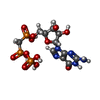
| Macromolecule | Name: PHOSPHOMETHYLPHOSPHONIC ACID GUANYLATE ESTER / type: ligand / ID: 2 / Number of copies: 4 / Formula: G2P |
|---|---|
| Molecular weight | Theoretical: 521.208 Da |
| Chemical component information |  ChemComp-G2P: |
-Experimental details
-Structure determination
| Method | cryo EM |
|---|---|
 Processing Processing | helical reconstruction |
| Aggregation state | filament |
- Sample preparation
| Concentration | 0.4 mg/mL |
|---|---|
| Buffer | pH: 7.4 |
| Grid | Model: Quantifoil R1.2/1.3 / Material: GOLD / Mesh: 300 / Support film - Material: CARBON / Support film - topology: HOLEY / Pretreatment - Type: GLOW DISCHARGE / Pretreatment - Time: 60 sec. / Pretreatment - Atmosphere: AIR |
| Vitrification | Cryogen name: ETHANE / Chamber humidity: 100 % / Chamber temperature: 289 K / Instrument: FEI VITROBOT MARK IV |
- Electron microscopy
| Microscope | TFS KRIOS |
|---|---|
| Image recording | Film or detector model: FEI FALCON III (4k x 4k) / Detector mode: COUNTING / Digitization - Dimensions - Width: 4096 pixel / Digitization - Dimensions - Height: 4096 pixel / Number grids imaged: 2 / Number real images: 3110 / Average exposure time: 60.0 sec. / Average electron dose: 23.5 e/Å2 |
| Electron beam | Acceleration voltage: 300 kV / Electron source:  FIELD EMISSION GUN FIELD EMISSION GUN |
| Electron optics | C2 aperture diameter: 50.0 µm / Calibrated magnification: 130841 / Illumination mode: FLOOD BEAM / Imaging mode: BRIGHT FIELD / Cs: 2.7 mm / Nominal defocus max: 3.0 µm / Nominal defocus min: 1.8 µm / Nominal magnification: 75000 |
| Sample stage | Specimen holder model: FEI TITAN KRIOS AUTOGRID HOLDER / Cooling holder cryogen: NITROGEN |
| Experimental equipment |  Model: Titan Krios / Image courtesy: FEI Company |
+ Image processing
-Atomic model buiding 1
| Initial model | Chain - Source name: AlphaFold / Chain - Initial model type: in silico model |
|---|---|
| Refinement | Space: REAL / Protocol: OTHER |
| Output model |  PDB-9v7v: |
 Movie
Movie Controller


 Z (Sec.)
 Y (Row.)
 X (Col.)


