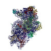
Movie
Movie Controller


+ Open data

- Basic information
| Entry |  | |||||||||
|---|---|---|---|---|---|---|---|---|---|---|
| Title | EMCV IRES captured on mammalian 40S with initiator tRNA | |||||||||
 Map data Map data | ||||||||||
 Sample Sample |
| |||||||||
 Keywords Keywords | EMCV IRES / 40S ribosome / 48S PIC / RIBOSOME | |||||||||
| Function / homology |  Function and homology information Function and homology informationlaminin receptor activity / ubiquitin ligase inhibitor activity / 90S preribosome / positive regulation of signal transduction by p53 class mediator / phagocytic cup / translation regulator activity / ribosomal small subunit export from nucleus / rough endoplasmic reticulum / laminin binding / positive regulation of apoptotic signaling pathway ...laminin receptor activity / ubiquitin ligase inhibitor activity / 90S preribosome / positive regulation of signal transduction by p53 class mediator / phagocytic cup / translation regulator activity / ribosomal small subunit export from nucleus / rough endoplasmic reticulum / laminin binding / positive regulation of apoptotic signaling pathway / MDM2/MDM4 family protein binding / DNA-(apurinic or apyrimidinic site) lyase / class I DNA-(apurinic or apyrimidinic site) endonuclease activity / maturation of SSU-rRNA from tricistronic rRNA transcript (SSU-rRNA, 5.8S rRNA, LSU-rRNA) / maturation of SSU-rRNA / small-subunit processome / spindle / rRNA processing / cytosolic ribosome / glucose homeostasis / ribosomal small subunit assembly / ribosome binding / ribosomal small subunit biogenesis / small ribosomal subunit rRNA binding / small ribosomal subunit / cytosolic small ribosomal subunit / perikaryon / cytoplasmic translation / rRNA binding / mitochondrial inner membrane / structural constituent of ribosome / ribosome / translation / mRNA binding / centrosome / nucleolus / dendrite / synapse / perinuclear region of cytoplasm / RNA binding / zinc ion binding / nucleus / plasma membrane / cytoplasm Similarity search - Function | |||||||||
| Biological species |   Oryctolagus cuniculus (rabbit) / Oryctolagus cuniculus (rabbit) /  Encephalomyocarditis virus Encephalomyocarditis virus | |||||||||
| Method | single particle reconstruction / cryo EM / Resolution: 4.6 Å | |||||||||
 Authors Authors | Das D / Hussain T | |||||||||
| Funding support |  India, 1 items India, 1 items
| |||||||||
 Citation Citation | Journal: Elife / Year: 2026 Title: Structural insights into the recruitment of viral type 2 IRES to ribosomal preinitiation complex for protein synthesis. Authors: Deepakash Das / Tanweer Hussain / Abstract: Picornaviruses employ internal ribosome entry sites (IRESs) in their genomic RNA to hijack the host's translational machinery. The picornavirus, encephalomyocarditis virus, employs a type 2 IRES ...Picornaviruses employ internal ribosome entry sites (IRESs) in their genomic RNA to hijack the host's translational machinery. The picornavirus, encephalomyocarditis virus, employs a type 2 IRES present in its 5' untranslated region (5'UTR) and requires 43S ribosomal preinitiation complex (PIC), the central domain of eukaryotic initiation factor (eIF) 4G, eIF4A, and an essential ITAF (IRES trans-acting factor)-polypyrimidine tract binding protein 1 (PTB1) to form 48S PIC. In this study, we have used cryo-electron microscopy (cryo-EM) to determine the structure of encephalomyocarditis virus (EMCV) IRES-bound mammalian 48S PIC in a scanning-arrested closed state at the start codon. The EMCV IRES domains contact initiator tRNA (tRNA) and 40S head at the inter-subunit interface, which reveals an altogether unique mechanism used by viruses to capture host translational machinery for its protein synthesis. The tRNA is held away from the 40S body in contrast to canonical cap-dependent translation while the domain I apical region of EMCV IRES mimics 28S rRNA of 60S to interact with 40S ribosomal head proteins uS13 and uS19. The structural analysis accounts for numerous previously reported biochemical studies on type 2 IRES and shows how type 2 IRES interacts with 43S PIC to form 48S PIC. This study provides mechanistic insights for understanding EMCV IRES-mediated translation initiation, which could be extrapolated to other IRESs sharing similar motifs and factor requirements, including type 1 viral IRESs. #1: Journal: Elife / Year: 2026Title: Structural insights into the recruitment of viral Type 2 IRES to ribosomal preinitiation complex for protein synthesis Authors: Das D / Hussain T | |||||||||
| History |
|
- Structure visualization
| Supplemental images |
|---|

- Downloads & links
-EMDB archive
| Map data | emd_64644.map.gz | 230.2 MB | EMDB map data format | |
|---|---|---|---|---|
| Header (meta data) | emd-64644-v30.xmlemd-64644.xml | 57.5 KB 57.5 KB | Display Display | EMDB header |
| FSC (resolution estimation) | emd_64644_fsc.xml | 13.3 KB | Display | FSC data file |
| Images |  emd_64644.png emd_64644.png | 54.2 KB | ||
| Filedesc metadata | emd-64644.cif.gz | 12.7 KB | ||
| Others | emd_64644_half_map_1.map.gzemd_64644_half_map_2.map.gz | 226.3 MB 226.3 MB | ||
| Archive directory |  https://data.pdbj.org/pub/emdb/structures/EMD-64644ftp://data.pdbj.org/pub/emdb/structures/EMD-64644 https://data.pdbj.org/pub/emdb/structures/EMD-64644ftp://data.pdbj.org/pub/emdb/structures/EMD-64644 | HTTPS FTP |
-Related structure data
| Related structure data |  9uzkMC  9uzlC  9uzmC M: atomic model generated by this map C: citing same article ( |
|---|---|
| Similar structure data |


-Links
| EMDB pages | EMDB (EBI/PDBe) / EMDataResource |
|---|---|
| Related items in Molecule of the Month |
-Map
| File | Download / File: emd_64644.map.gz / Format: CCP4 / Size: 244.1 MB / Type: IMAGE STORED AS FLOATING POINT NUMBER (4 BYTES) | ||||||||||||||||||||||||||||||||||||
|---|---|---|---|---|---|---|---|---|---|---|---|---|---|---|---|---|---|---|---|---|---|---|---|---|---|---|---|---|---|---|---|---|---|---|---|---|---|
| Projections & slices | Image control
Images are generated by Spider. | ||||||||||||||||||||||||||||||||||||
| Voxel size | X=Y=Z: 1.17 Å | ||||||||||||||||||||||||||||||||||||
| Density |
| ||||||||||||||||||||||||||||||||||||
| Symmetry | Space group: 1 | ||||||||||||||||||||||||||||||||||||
| Details | EMDB XML:
|
 Z (Sec.)
 Y (Row.)
 X (Col.)


-Supplemental data
-Half map: #2
| File | emd_64644_half_map_1.map | ||||||||||||
|---|---|---|---|---|---|---|---|---|---|---|---|---|---|
| Projections & Slices |
| ||||||||||||
| Density Histograms |


-Half map: #1
| File | emd_64644_half_map_2.map | ||||||||||||
|---|---|---|---|---|---|---|---|---|---|---|---|---|---|
| Projections & Slices |
| ||||||||||||
| Density Histograms |


- Sample components
+Entire : EMCV IRES with 40S and initiator tRNA
+Supramolecule #1: EMCV IRES with 40S and initiator tRNA
+Macromolecule #1: 18S ribosomal RNA
+Macromolecule #36: Initiator tRNA
+Macromolecule #37: EMCV IRES
+Macromolecule #2: Small ribosomal subunit protein uS2
+Macromolecule #3: Small ribosomal subunit protein eS1
+Macromolecule #4: Small ribosomal subunit protein uS5
+Macromolecule #5: Small ribosomal subunit protein uS3
+Macromolecule #6: Small ribosomal subunit protein eS4
+Macromolecule #7: Small ribosomal subunit protein uS7
+Macromolecule #8: Small ribosomal subunit protein eS6
+Macromolecule #9: Small ribosomal subunit protein eS7
+Macromolecule #10: Small ribosomal subunit protein eS8
+Macromolecule #11: Small ribosomal subunit protein uS4
+Macromolecule #12: Small ribosomal subunit protein eS10
+Macromolecule #13: Small ribosomal subunit protein uS17
+Macromolecule #14: Small ribosomal subunit protein eS12
+Macromolecule #15: Small ribosomal subunit protein uS15
+Macromolecule #16: Ribosomal protein S14
+Macromolecule #17: Small ribosomal subunit protein uS19
+Macromolecule #18: Small ribosomal subunit protein uS9
+Macromolecule #19: Small ribosomal subunit protein eS17
+Macromolecule #20: Small ribosomal subunit protein uS13
+Macromolecule #21: Small ribosomal subunit protein eS19
+Macromolecule #22: Small ribosomal subunit protein uS10
+Macromolecule #23: Small ribosomal subunit protein eS21
+Macromolecule #24: Small ribosomal subunit protein uS8
+Macromolecule #25: Small ribosomal subunit protein uS12
+Macromolecule #26: 40S ribosomal protein S24
+Macromolecule #27: Small ribosomal subunit protein eS26
+Macromolecule #28: Small ribosomal subunit protein eS27
+Macromolecule #29: Small ribosomal subunit protein eS28
+Macromolecule #30: Small ribosomal subunit protein uS14
+Macromolecule #31: Ubiquitin-ribosomal protein eS31 fusion protein
+Macromolecule #32: Small ribosomal subunit protein RACK1
+Macromolecule #33: Small ribosomal subunit protein eS25
+Macromolecule #34: Small ribosomal subunit protein eS30
+Macromolecule #35: Small ribosomal subunit protein eS32
-Experimental details
-Structure determination
| Method | cryo EM |
|---|---|
 Processing Processing | single particle reconstruction |
| Aggregation state | particle |
-Sample preparation
| Buffer | pH: 7.4 Details: 20mM HEPES, 150mM KOAc, 2mM MgCl2, 1mM DTT, 0.5mM Spermidine, 6mM GMP-PnP |
|---|---|
| Grid | Model: C-flat-1.2/1.3 / Material: COPPER / Mesh: 400 / Support film - Material: CARBON / Support film - topology: CONTINUOUS / Pretreatment - Type: GLOW DISCHARGE / Pretreatment - Time: 70 sec. / Pretreatment - Atmosphere: OTHER |
| Vitrification | Cryogen name: ETHANE-PROPANE / Chamber humidity: 100 % / Chamber temperature: 289 K / Instrument: FEI VITROBOT MARK IV |
- Electron microscopy
| Microscope | FEI TALOS ARCTICA |
|---|---|
| Temperature | Min: 77.0 K / Max: 83.0 K |
| Image recording | Film or detector model: GATAN K2 SUMMIT (4k x 4k) / Detector mode: COUNTING / Digitization - Dimensions - Width: 3838 pixel / Digitization - Dimensions - Height: 3710 pixel / Number grids imaged: 6 / Number real images: 22549 / Average electron dose: 55.0 e/Å2 |
| Electron beam | Acceleration voltage: 200 kV / Electron source:  FIELD EMISSION GUN FIELD EMISSION GUN |
| Electron optics | C2 aperture diameter: 70.0 µm / Illumination mode: OTHER / Imaging mode: BRIGHT FIELD / Cs: 2.7 mm / Nominal defocus max: 2.75 µm / Nominal defocus min: 1.25 µm / Nominal magnification: 36000 |
| Sample stage | Specimen holder model: OTHER / Cooling holder cryogen: NITROGEN |
| Experimental equipment | 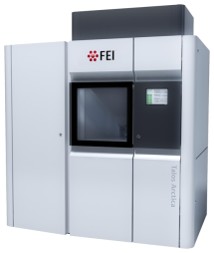 Model: Talos Arctica / Image courtesy: FEI Company |
+Image processing

-Atomic model buiding 1
| Initial model |
| ||||||||
|---|---|---|---|---|---|---|---|---|---|
| Output model | PDB-9uzk: |