 Movie
Movie Controller


+ Open data

- Basic information
| Entry |  | |||||||||
|---|---|---|---|---|---|---|---|---|---|---|
| Title | Cryo-EM structure of the ZQ16-bound GPR84 receptor-Gi complex | |||||||||
 Map data Map data | Cryo-EM structure of the ZQ-16-bound GPR84 receptor-Gi complex | |||||||||
 Sample Sample |
| |||||||||
 Keywords Keywords | GPCR / MCFA / complex / agonist / MEMBRANE PROTEIN | |||||||||
| Function / homology |  Function and homology information Function and homology informationurotensin II receptor activity / tertiary granule membrane / specific granule membrane / regulation of eating behavior / adenylate cyclase inhibitor activity / T cell migration / positive regulation of protein localization to cell cortex / positive regulation of relaxation of smooth muscle / Adenylate cyclase inhibitory pathway / D2 dopamine receptor binding ...urotensin II receptor activity / tertiary granule membrane / specific granule membrane / regulation of eating behavior / adenylate cyclase inhibitor activity / T cell migration / positive regulation of protein localization to cell cortex / positive regulation of relaxation of smooth muscle / Adenylate cyclase inhibitory pathway / D2 dopamine receptor binding / adenylate cyclase-inhibiting dopamine receptor signaling pathway / adenylate cyclase-inhibiting serotonin receptor signaling pathway / G protein-coupled serotonin receptor binding / regulation of G protein-coupled receptor signaling pathway / cellular response to forskolin / regulation of mitotic spindle organization / mast cell degranulation / chemokine-mediated signaling pathway / establishment of mitotic spindle orientation / neuropeptide signaling pathway / Regulation of insulin secretion / response to prostaglandin E / positive regulation of cholesterol biosynthetic process / G protein-coupled receptor binding / response to peptide hormone / G-protein beta/gamma-subunit complex binding / adenylate cyclase-inhibiting G protein-coupled receptor signaling pathway / adenylate cyclase-modulating G protein-coupled receptor signaling pathway / Olfactory Signaling Pathway / Activation of the phototransduction cascade / G protein-coupled acetylcholine receptor signaling pathway / G beta:gamma signalling through PLC beta / Presynaptic function of Kainate receptors / Thromboxane signalling through TP receptor / Activation of G protein gated Potassium channels / Inhibition of voltage gated Ca2+ channels via Gbeta/gamma subunits / G-protein activation / Glucagon signaling in metabolic regulation / G beta:gamma signalling through CDC42 / Prostacyclin signalling through prostacyclin receptor / Synthesis, secretion, and inactivation of Glucagon-like Peptide-1 (GLP-1) / GDP binding / photoreceptor disc membrane / G beta:gamma signalling through BTK / ADP signalling through P2Y purinoceptor 12 / Sensory perception of sweet, bitter, and umami (glutamate) taste / Glucagon-type ligand receptors / Adrenaline,noradrenaline inhibits insulin secretion / Vasopressin regulates renal water homeostasis via Aquaporins / G alpha (z) signalling events / Glucagon-like Peptide-1 (GLP1) regulates insulin secretion / cellular response to catecholamine stimulus / ADP signalling through P2Y purinoceptor 1 / G beta:gamma signalling through PI3Kgamma / ADORA2B mediated anti-inflammatory cytokines production / adenylate cyclase-activating dopamine receptor signaling pathway / cellular response to prostaglandin E stimulus / Cooperation of PDCL (PhLP1) and TRiC/CCT in G-protein beta folding / GPER1 signaling / heterotrimeric G-protein complex / G alpha (12/13) signalling events / Inactivation, recovery and regulation of the phototransduction cascade / G-protein beta-subunit binding / extracellular vesicle / Thrombin signalling through proteinase activated receptors (PARs) / adenylate cyclase-activating G protein-coupled receptor signaling pathway / signaling receptor complex adaptor activity / spindle pole / ciliary basal body / GTPase binding / midbody / G protein activity / Ca2+ pathway / cell cortex / High laminar flow shear stress activates signaling by PIEZO1 and PECAM1:CDH5:KDR in endothelial cells / G alpha (i) signalling events / G alpha (s) signalling events / G alpha (q) signalling events / Hydrolases; Acting on acid anhydrides; Acting on GTP to facilitate cellular and subcellular movement / cytoplasmic side of plasma membrane / Ras protein signal transduction / cell division / signaling receptor complex / Extra-nuclear estrogen signaling / G protein-coupled receptor signaling pathway / lysosomal membrane / centrosome / GTPase activity / Neutrophil degranulation / GTP binding / synapse / protein-containing complex binding / magnesium ion binding / signal transduction / extracellular exosome / membrane / nucleus / plasma membrane / cytosol / cytoplasm Similarity search - Function | |||||||||
| Biological species |  Homo sapiens (human) Homo sapiens (human) | |||||||||
| Method | single particle reconstruction / cryo EM / Resolution: 3.1 Å | |||||||||
 Authors Authors | Choi MK / Cho HS | |||||||||
| Funding support | 1 items
| |||||||||
 Citation Citation | Journal: Exp Mol Med / Year: 2026 Title: Pharmacological modulation of GPR84 revealed by dual states structures and immune functional assays. Authors: Myung Kyung Choi / Dong Jin Park / Pankyung Kim / Hee Seong Choi / Sorin Myung / Youngki Yoo / Nienping Chang / Ga-Yeon Yoon / Hye Jin Kang / Sang-Jun Ha / Hyun-Soo Cho /   Abstract: G-protein-coupled receptor 84 (GPR84) is an orphan class A GPCR selectively activated by medium chain fatty acids and highly expressed in immune cells, where it modulates pro-inflammatory signaling. ...G-protein-coupled receptor 84 (GPR84) is an orphan class A GPCR selectively activated by medium chain fatty acids and highly expressed in immune cells, where it modulates pro-inflammatory signaling. The structural basis of GPR84 inactivation and antagonism has remained unclear, limiting the rational design of pathway-selective modulators despite its clinical relevance in metabolic inflammation and fibrotic diseases. Here, we report cryo-electron microscopy structures of human GPR84 in inactive and active states. The 3.5 Å inactive structure bound to the antagonist GLPG1205 reveals a lid-like conformation of extracellular loop 2 and an inward reorientation of Arg172, with the antagonist head group blocking the allosteric sodium-binding site. Molecular dynamics simulations further support these findings, identifying an aberrant TM5, TM6 lateral entry gate. By contrast, the 3.17 Å agonist ZQ-16, Gαi complex, shows a rearranged toggle switch and comparative analyses highlight extracellular loop 2 conformational plasticity. Immune functional assays in THP-1 cells demonstrated that ZQ-16 elicited GPR84-dependent activation and cytokine production, which were effectively abrogated by GLPG1205. Mutagenesis combined with functional assays validates key ligand interactions, providing a framework for the rational design of pathway selective GPR84 modulators. | |||||||||
| History |
|
- Structure visualization
| Supplemental images |
|---|

- Downloads & links
-EMDB archive
| Map data | emd_64407.map.gz | 81.7 MB | EMDB map data format | |
|---|---|---|---|---|
| Header (meta data) | emd-64407-v30.xmlemd-64407.xml | 22 KB 22 KB | Display Display | EMDB header |
| FSC (resolution estimation) | emd_64407_fsc.xml | 16 KB | Display | FSC data file |
| Images |  emd_64407.png emd_64407.png | 23.1 KB | ||
| Filedesc metadata | emd-64407.cif.gz | 6.9 KB | ||
| Others | emd_64407_half_map_1.map.gzemd_64407_half_map_2.map.gz | 151.8 MB 151.8 MB | ||
| Archive directory |  https://data.pdbj.org/pub/emdb/structures/EMD-64407ftp://data.pdbj.org/pub/emdb/structures/EMD-64407 https://data.pdbj.org/pub/emdb/structures/EMD-64407ftp://data.pdbj.org/pub/emdb/structures/EMD-64407 | HTTPS FTP |
-Related structure data
| Related structure data |  9upyMC  9v6tC M: atomic model generated by this map C: citing same article ( |
|---|---|
| Similar structure data |

-Links
| EMDB pages | EMDB (EBI/PDBe) / EMDataResource |
|---|---|
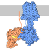
| Related items in Molecule of the Month |





















-Map
| File | Download / File: emd_64407.map.gz / Format: CCP4 / Size: 163.6 MB / Type: IMAGE STORED AS FLOATING POINT NUMBER (4 BYTES) | ||||||||||||||||||||||||||||||||||||
|---|---|---|---|---|---|---|---|---|---|---|---|---|---|---|---|---|---|---|---|---|---|---|---|---|---|---|---|---|---|---|---|---|---|---|---|---|---|
| Annotation | Cryo-EM structure of the ZQ-16-bound GPR84 receptor-Gi complex | ||||||||||||||||||||||||||||||||||||
| Projections & slices | Image control
Images are generated by Spider. | ||||||||||||||||||||||||||||||||||||
| Voxel size | X=Y=Z: 0.9013 Å | ||||||||||||||||||||||||||||||||||||
| Density |
| ||||||||||||||||||||||||||||||||||||
| Symmetry | Space group: 1 | ||||||||||||||||||||||||||||||||||||
| Details | EMDB XML:
|
 Z (Sec.)
 Y (Row.)
 X (Col.)


-Supplemental data
-Half map: Half B map
| File | emd_64407_half_map_1.map | ||||||||||||
|---|---|---|---|---|---|---|---|---|---|---|---|---|---|
| Annotation | Half_B map | ||||||||||||
| Projections & Slices |
| ||||||||||||
| Density Histograms |


-Half map: Half A map
| File | emd_64407_half_map_2.map | ||||||||||||
|---|---|---|---|---|---|---|---|---|---|---|---|---|---|
| Annotation | Half_A map | ||||||||||||
| Projections & Slices |
| ||||||||||||
| Density Histograms |


- Sample components
-Entire : ZQ16-bound GPR84 receptor-Gi complex
| Entire | Name: ZQ16-bound GPR84 receptor-Gi complex |
|---|---|
| Components |
|
-Supramolecule #1: ZQ16-bound GPR84 receptor-Gi complex
| Supramolecule | Name: ZQ16-bound GPR84 receptor-Gi complex / type: complex / ID: 1 / Parent: 0 / Macromolecule list: #1-#4 |
|---|---|
| Source (natural) | Organism: Homo sapiens (human) |
| Molecular weight | Theoretical: 130 KDa |
-Macromolecule #1: G-protein coupled receptor 84
| Macromolecule | Name: G-protein coupled receptor 84 / type: protein_or_peptide / ID: 1 / Number of copies: 1 / Enantiomer: LEVO |
|---|---|
| Source (natural) | Organism: Homo sapiens (human) |
| Molecular weight | Theoretical: 43.537535 KDa |
| Recombinant expression | Organism:   Spodoptera frugiperda (fall armyworm) Spodoptera frugiperda (fall armyworm) |
| Sequence | String: DYKDDDDKSS DANFSCYHES VLGYRYVAVS WGVVVAVTGT VGNVLTLLAL AIQPKLRTRF NLLIANLTLA DLLYCTLLQP FSVDTYLHL HWRTGATFCR VFGLLLFASN SVSILTLCLI ALGRYLLIAH PKLFPQVFSA KGIVLALVST WVVGVASFAP L WPIYILVP ...String: DYKDDDDKSS DANFSCYHES VLGYRYVAVS WGVVVAVTGT VGNVLTLLAL AIQPKLRTRF NLLIANLTLA DLLYCTLLQP FSVDTYLHL HWRTGATFCR VFGLLLFASN SVSILTLCLI ALGRYLLIAH PKLFPQVFSA KGIVLALVST WVVGVASFAP L WPIYILVP VVCTCSFDRI RGRPYTTILM GIYFVLGLSS VGIFYCLIHR QVKRAAQALD QYKLRQASIH SNHVARTDEA MP GRFQELD SRLASGGPSE GISSEPVSAA TTQTLEGDSS EVGDQINSKR AKQMAEKSPP EASAKAQPIK GARRAPDSSS EFG KVTRMC FAVFLCFALS YIPFLLLNIL DARVQAPRVV HMLAANLTWL NGCINPVLYA AMNRQFRQAY GSILKRGPR UniProtKB: G protein-coupled receptor 84 |
-Macromolecule #2: Guanine nucleotide-binding protein G(i) subunit alpha-1
| Macromolecule | Name: Guanine nucleotide-binding protein G(i) subunit alpha-1 type: protein_or_peptide / ID: 2 / Number of copies: 1 / Enantiomer: LEVO EC number: Hydrolases; Acting on acid anhydrides; Acting on GTP to facilitate cellular and subcellular movement |
|---|---|
| Source (natural) | Organism: Homo sapiens (human) |
| Molecular weight | Theoretical: 40.691316 KDa |
| Recombinant expression | Organism: Spodoptera frugiperda (fall armyworm) |
| Sequence | String: EFMGCTLSAE DKAAVERSKM IDRNLREDGE KAAREVKLLL LGAGESGKST IVKQMKIIHE AGYSEEECKQ YKAVVYSNTI QSIIAIIRA MGRLKIDFGD SARADDARQL FVLAGAAEEG FMTAELAGVI KRLWKDSGVQ ACFNRSREYQ LNDSAAYYLN D LDRIAQPN ...String: EFMGCTLSAE DKAAVERSKM IDRNLREDGE KAAREVKLLL LGAGESGKST IVKQMKIIHE AGYSEEECKQ YKAVVYSNTI QSIIAIIRA MGRLKIDFGD SARADDARQL FVLAGAAEEG FMTAELAGVI KRLWKDSGVQ ACFNRSREYQ LNDSAAYYLN D LDRIAQPN YIPTQQDVLR TRVKTTGIVE THFTFKDLHF KMFDVGGQRS ERKKWIHCFE GVTAIIFCVA LSDYDLVLAE DE EMNRMHE SMKLFDSICN NKWFTDTSII LFLNKKDLFE EKIKKSPLTI CYPEYAGSNT YEEAAAYIQC QFEDLNKRKD TKE IYTHFT CATDTKNVQF VFDAVTDVII KNNLKDCGLF UniProtKB: Guanine nucleotide-binding protein G(i) subunit alpha-1 |
-Macromolecule #3: Guanine nucleotide-binding protein G(I)/G(S)/G(T) subunit beta-1
| Macromolecule | Name: Guanine nucleotide-binding protein G(I)/G(S)/G(T) subunit beta-1 type: protein_or_peptide / ID: 3 / Number of copies: 1 / Enantiomer: LEVO |
|---|---|
| Source (natural) | Organism: Homo sapiens (human) |
| Molecular weight | Theoretical: 39.475137 KDa |
| Recombinant expression | Organism: Spodoptera frugiperda (fall armyworm) |
| Sequence | String: MHHHHHHLEV LFQGPGSSGS ELDQLRQEAE QLKNQIRDAR KACADATLSQ ITNNIDPVGR IQMRTRRTLR GHLAKIYAMH WGTDSRLLV SASQDGKLII WDSYTTNKVH AIPLRSSWVM TCAYAPSGNY VACGGLDNIC SIYNLKTREG NVRVSRELAG H TGYLSCCR ...String: MHHHHHHLEV LFQGPGSSGS ELDQLRQEAE QLKNQIRDAR KACADATLSQ ITNNIDPVGR IQMRTRRTLR GHLAKIYAMH WGTDSRLLV SASQDGKLII WDSYTTNKVH AIPLRSSWVM TCAYAPSGNY VACGGLDNIC SIYNLKTREG NVRVSRELAG H TGYLSCCR FLDDNQIVTS SGDTTCALWD IETGQQTTTF TGHTGDVMSL SLAPDTRLFV SGACDASAKL WDVREGMCRQ TF TGHESDI NAICFFPNGN AFATGSDDAT CRLFDLRADQ ELMTYSHDNI ICGITSVSFS KSGRLLLAGY DDFNCNVWDA LKA DRAGVL AGHDNRVSCL GVTDDGMAVA TGSWDSFLKI WNG UniProtKB: Guanine nucleotide-binding protein G(I)/G(S)/G(T) subunit beta-1 |
-Macromolecule #4: Guanine nucleotide-binding protein G(I)/G(S)/G(O) subunit gamma-2
| Macromolecule | Name: Guanine nucleotide-binding protein G(I)/G(S)/G(O) subunit gamma-2 type: protein_or_peptide / ID: 4 / Number of copies: 1 / Enantiomer: LEVO |
|---|---|
| Source (natural) | Organism: Homo sapiens (human) |
| Molecular weight | Theoretical: 7.861143 KDa |
| Recombinant expression | Organism: Spodoptera frugiperda (fall armyworm) |
| Sequence | String: MASNNTASIA QARKLVEQLK MEANIDRIKV SKAAADLMAY CEAHAKEDPL LTPVPASENP FREKKFFCAI L UniProtKB: Guanine nucleotide-binding protein G(I)/G(S)/G(O) subunit gamma-2 |
-Macromolecule #5: 2-hexylsulfanyl-4-oxidanyl-1~{H}-pyrimidin-6-one
| Macromolecule | Name: 2-hexylsulfanyl-4-oxidanyl-1~{H}-pyrimidin-6-one / type: ligand / ID: 5 / Number of copies: 1 / Formula: A1L9K |
|---|---|
| Molecular weight | Theoretical: 228.311 Da |
-Experimental details
-Structure determination
| Method | cryo EM |
|---|---|
 Processing Processing | single particle reconstruction |
| Aggregation state | particle |
-Sample preparation
| Concentration | 3 mg/mL | |||||||||||||||||||||
|---|---|---|---|---|---|---|---|---|---|---|---|---|---|---|---|---|---|---|---|---|---|---|
| Buffer | pH: 7.5 Component:
| |||||||||||||||||||||
| Grid | Model: Quantifoil / Material: COPPER / Mesh: 300 / Support film - Material: CARBON / Support film - topology: HOLEY / Pretreatment - Type: GLOW DISCHARGE | |||||||||||||||||||||
| Vitrification | Cryogen name: ETHANE / Chamber humidity: 100 % / Chamber temperature: 277.15 K / Instrument: FEI VITROBOT MARK IV |
- Electron microscopy
| Microscope | TFS KRIOS |
|---|---|
| Image recording | Film or detector model: FEI FALCON IV (4k x 4k) / Average electron dose: 50.0 e/Å2 |
| Electron beam | Acceleration voltage: 300 kV / Electron source:  FIELD EMISSION GUN FIELD EMISSION GUN |
| Electron optics | Illumination mode: FLOOD BEAM / Imaging mode: BRIGHT FIELD / Nominal defocus max: 2.1 µm / Nominal defocus min: 0.8 µm |
| Sample stage | Specimen holder model: FEI TITAN KRIOS AUTOGRID HOLDER / Cooling holder cryogen: NITROGEN |
| Experimental equipment |  Model: Titan Krios / Image courtesy: FEI Company |

