[English] 日本語
 Yorodumi
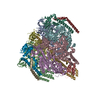
- EMDB-63929: Cryo-EM structure of the Vo domain of V/A-ATPase in liposomes und... -
+ Open data
- Basic information
| Entry |  | ||||||||||||
|---|---|---|---|---|---|---|---|---|---|---|---|---|---|
| Title | Cryo-EM structure of the Vo domain of V/A-ATPase in liposomes under pmf condition,state1 | ||||||||||||
 Map data Map data | |||||||||||||
 Sample Sample |
| ||||||||||||
 Keywords Keywords | ATP synthase / V ATPase / V1 ATPase / MOTOR PROTEIN | ||||||||||||
| Biological species |   Thermus thermophilus HB8 (bacteria) Thermus thermophilus HB8 (bacteria) | ||||||||||||
| Method | single particle reconstruction / cryo EM / Resolution: 3.2 Å | ||||||||||||
 Authors Authors | Nakano A / Kishikawa J / Nishida Y / Gerle C / Shigematsu H / Mitsuoka M / Yokoyama K | ||||||||||||
| Funding support |  Japan, 3 items Japan, 3 items
| ||||||||||||
 Citation Citation |  Journal: Sci Adv / Year: 2025 Journal: Sci Adv / Year: 2025Title: Structures of rotary ATP synthase from during proton powered ATP synthesis. Authors: Atsuki Nakano / Jun-Ichi Kishikawa / Nishida Yui / Kyosuke Sugawara / Yuto Kan / Christoph Gerle / Hideki Shigematsu / Kaoru Mitsuoka / Ken Yokoyama /  Abstract: ATP synthases are rotary molecular machines that use the proton motive force to rotate the central rotor complex relative to the surrounding stator apparatus, thereby coupling the ATP synthesis. We ...ATP synthases are rotary molecular machines that use the proton motive force to rotate the central rotor complex relative to the surrounding stator apparatus, thereby coupling the ATP synthesis. We reconstituted the V/A-ATPase into liposomes and performed structural analysis using cryo-EM under conditions where the proton motive force was applied in the presence of ADP and Pi. ATP molecules were bound at two of the three catalytic sites of V/A-ATPase, confirming that the structure represents a state adopted during ATP synthesis. In this structure, the catalytic site closes upon binding of ADP and Pi through an induced fit mechanism. Multiple structures were obtained where the membrane-embedded rotor ring was in a different position relative to the stator. By comparing these structures, we found that torsion occurs in both the central rotor and the peripheral stator during 31° rotation of rotor ring. These structural snapshots of V/A-ATPase provide crucial insights into the mechanism of rotary catalysis of ATP synthesis. | ||||||||||||
| History |
|
- Structure visualization
| Supplemental images |
|---|
- Downloads & links
-EMDB archive
| Map data |  emd_63929.map.gz emd_63929.map.gz | 96.2 MB |  EMDB map data format EMDB map data format | |
|---|---|---|---|---|
| Header (meta data) |  emd-63929-v30.xml emd-63929-v30.xml emd-63929.xml emd-63929.xml | 18.1 KB 18.1 KB | Display Display |  EMDB header EMDB header |
| FSC (resolution estimation) |  emd_63929_fsc.xml emd_63929_fsc.xml | 11.4 KB | Display |  FSC data file FSC data file |
| Images |  emd_63929.png emd_63929.png | 71.5 KB | ||
| Masks |  emd_63929_msk_1.map emd_63929_msk_1.map | 125 MB |  Mask map Mask map | |
| Filedesc metadata |  emd-63929.cif.gz emd-63929.cif.gz | 4.5 KB | ||
| Others |  emd_63929_additional_1.map.gz emd_63929_additional_1.map.gz emd_63929_half_map_1.map.gz emd_63929_half_map_1.map.gz emd_63929_half_map_2.map.gz emd_63929_half_map_2.map.gz | 115.2 MB 97.5 MB 97.4 MB | ||
| Archive directory |  http://ftp.pdbj.org/pub/emdb/structures/EMD-63929 http://ftp.pdbj.org/pub/emdb/structures/EMD-63929 ftp://ftp.pdbj.org/pub/emdb/structures/EMD-63929 ftp://ftp.pdbj.org/pub/emdb/structures/EMD-63929 | HTTPS FTP |
-Related structure data
| Related structure data |  9u6fC  9u6gC  9u6hC  9u6iC  9u6jC  9u6kC  9u6lC  9u6mC  9u6nC  9u6oC  9u6pC  9u6qC  9u6tC  9u6uC  9u6vC  9u6wC  9u6xC  9u6yC C: citing same article ( |
|---|
- Links
Links
| EMDB pages |  EMDB (EBI/PDBe) / EMDB (EBI/PDBe) /  EMDataResource EMDataResource |
|---|
- Map
Map
| File |  Download / File: emd_63929.map.gz / Format: CCP4 / Size: 125 MB / Type: IMAGE STORED AS FLOATING POINT NUMBER (4 BYTES) Download / File: emd_63929.map.gz / Format: CCP4 / Size: 125 MB / Type: IMAGE STORED AS FLOATING POINT NUMBER (4 BYTES) | ||||||||||||||||||||||||||||||||||||
|---|---|---|---|---|---|---|---|---|---|---|---|---|---|---|---|---|---|---|---|---|---|---|---|---|---|---|---|---|---|---|---|---|---|---|---|---|---|
| Projections & slices | Image control
Images are generated by Spider. | ||||||||||||||||||||||||||||||||||||
| Voxel size | X=Y=Z: 1.05 Å | ||||||||||||||||||||||||||||||||||||
| Density |
| ||||||||||||||||||||||||||||||||||||
| Symmetry | Space group: 1 | ||||||||||||||||||||||||||||||||||||
| Details | EMDB XML:
|
-Supplemental data
-Mask #1
| File |  emd_63929_msk_1.map emd_63929_msk_1.map | ||||||||||||
|---|---|---|---|---|---|---|---|---|---|---|---|---|---|
| Projections & Slices |
| ||||||||||||
| Density Histograms |
-Additional map: #1
| File | emd_63929_additional_1.map | ||||||||||||
|---|---|---|---|---|---|---|---|---|---|---|---|---|---|
| Projections & Slices |
| ||||||||||||
| Density Histograms |
-Half map: #1
| File | emd_63929_half_map_1.map | ||||||||||||
|---|---|---|---|---|---|---|---|---|---|---|---|---|---|
| Projections & Slices |
| ||||||||||||
| Density Histograms |
-Half map: #2
| File | emd_63929_half_map_2.map | ||||||||||||
|---|---|---|---|---|---|---|---|---|---|---|---|---|---|
| Projections & Slices |
| ||||||||||||
| Density Histograms |
- Sample components
-Entire : Vo domain of V/A-ATPase from Thermus thermophilus
| Entire | Name: Vo domain of V/A-ATPase from Thermus thermophilus |
|---|---|
| Components |
|
-Supramolecule #1: Vo domain of V/A-ATPase from Thermus thermophilus
| Supramolecule | Name: Vo domain of V/A-ATPase from Thermus thermophilus / type: complex / ID: 1 / Parent: 0 / Macromolecule list: #1-#2 |
|---|---|
| Source (natural) | Organism:   Thermus thermophilus HB8 (bacteria) Thermus thermophilus HB8 (bacteria) |
| Molecular weight | Theoretical: 660 KDa |
-Experimental details
-Structure determination
| Method | cryo EM |
|---|---|
 Processing Processing | single particle reconstruction |
| Aggregation state | particle |
- Sample preparation
| Buffer | pH: 8 |
|---|---|
| Vitrification | Cryogen name: ETHANE / Chamber humidity: 100 % / Chamber temperature: 298 K |
- Electron microscopy
| Microscope | TFS KRIOS |
|---|---|
| Image recording | Film or detector model: GATAN K3 BIOQUANTUM (6k x 4k) / Average electron dose: 50.0 e/Å2 |
| Electron beam | Acceleration voltage: 300 kV / Electron source:  FIELD EMISSION GUN FIELD EMISSION GUN |
| Electron optics | Illumination mode: FLOOD BEAM / Imaging mode: BRIGHT FIELD / Cs: 2.7 mm / Nominal defocus max: 1.6 µm / Nominal defocus min: 1.4000000000000001 µm / Nominal magnification: 60000 |
| Sample stage | Cooling holder cryogen: NITROGEN |
| Experimental equipment |  Model: Titan Krios / Image courtesy: FEI Company |
+ Image processing
-Atomic model buiding 1
| Initial model | PDB ID: Chain - Source name: PDB / Chain - Initial model type: experimental model |
|---|---|
| Refinement | Space: REAL / Protocol: FLEXIBLE FIT |
 Movie
Movie Controller


 Z (Sec.)
 Y (Row.)
 X (Col.)


