 ムービー
ムービー コントローラー


+ データを開く

- 基本情報
基本情報
| 登録情報 | データベース: EMDB / ID: EMD-6360 | |||||||||
|---|---|---|---|---|---|---|---|---|---|---|
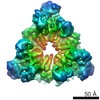
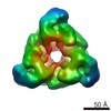
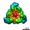
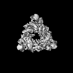
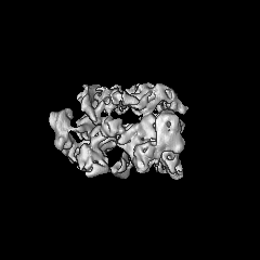
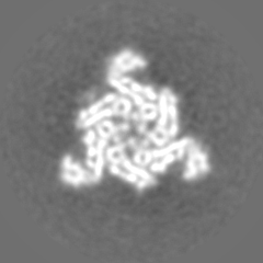
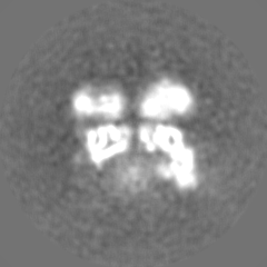
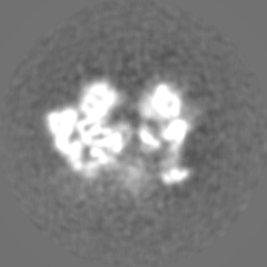
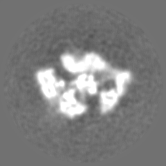
| タイトル | Cryo-EM structure of the peroxisomal Pex1/Pex6 complex in ADP state | |||||||||
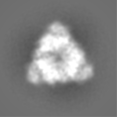
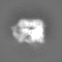
 マップデータ マップデータ | Reconstruction of Pex1/Pex6 AAA ATPase complex in ADP using single-particle cryo-electron microscopy without imposed symmetry | |||||||||
 試料 試料 |
| |||||||||
 キーワード キーワード | AAA ATPase / cryoelectron microscopy / peroxisome | |||||||||
| 機能・相同性 |  機能・相同性情報 機能・相同性情報protein import into peroxisome matrix, receptor recycling / protein import into peroxisome matrix / protein transporter activity / peroxisomal membrane / ATPase complex / protein unfolding / 加水分解酵素; 酸無水物に作用; 酸無水物に作用・細胞または細胞小器官の運動に関与 / peroxisome / ATP hydrolysis activity / ATP binding / cytosol 類似検索 - 分子機能 | |||||||||
| 生物種 |  | |||||||||
| 手法 | 単粒子再構成法 / クライオ電子顕微鏡法 / 解像度: 8.8 Å | |||||||||
 データ登録者 データ登録者 | Blok NB / Tan D / Wang RY / Penczek PA / Baker D / DiMaio F / Rapoport TA / Walz T | |||||||||
 引用 引用 | ジャーナル: Proc Natl Acad Sci U S A / 年: 2015 タイトル: Unique double-ring structure of the peroxisomal Pex1/Pex6 ATPase complex revealed by cryo-electron microscopy. 著者: Neil B Blok / Dongyan Tan / Ray Yu-Ruei Wang / Pawel A Penczek / David Baker / Frank DiMaio / Tom A Rapoport / Thomas Walz /  要旨: Members of the AAA family of ATPases assemble into hexameric double rings and perform vital functions, yet their molecular mechanisms remain poorly understood. Here, we report structures of the ...Members of the AAA family of ATPases assemble into hexameric double rings and perform vital functions, yet their molecular mechanisms remain poorly understood. Here, we report structures of the Pex1/Pex6 complex; mutations in these proteins frequently cause peroxisomal diseases. The structures were determined in the presence of different nucleotides by cryo-electron microscopy. Models were generated using a computational approach that combines Monte Carlo placement of structurally homologous domains into density maps with energy minimization and refinement protocols. Pex1 and Pex6 alternate in an unprecedented hexameric double ring. Each protein has two N-terminal domains, N1 and N2, structurally related to the single N domains in p97 and N-ethylmaleimide sensitive factor (NSF); N1 of Pex1 is mobile, but the others are packed against the double ring. The N-terminal ATPase domains are inactive, forming a symmetric D1 ring, whereas the C-terminal domains are active, likely in different nucleotide states, and form an asymmetric D2 ring. These results suggest how subunit activity is coordinated and indicate striking similarities between Pex1/Pex6 and p97, supporting the hypothesis that the Pex1/Pex6 complex has a role in peroxisomal protein import analogous to p97 in ER-associated protein degradation. | |||||||||
| 履歴 |
|
- 構造の表示
構造の表示
| ムービー |
ムービービューア |
|---|---|
| 構造ビューア | EMマップ: SurfViewMolmilJmol/JSmol |
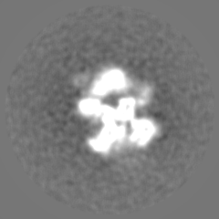
| 添付画像 |

- ダウンロードとリンク
-EMDBアーカイブ
| マップデータ | emd_6360.map.gz | 40 MB | EMDBマップデータ形式 | |
|---|---|---|---|---|
| ヘッダ (付随情報) | emd-6360-v30.xmlemd-6360.xml | 11.8 KB 11.8 KB | 表示 表示 | EMDBヘッダ |
| 画像 | 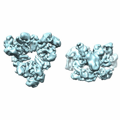 400_6360.gif 400_6360.gif 80_6360.gif 80_6360.gif | 49.9 KB 3.1 KB | ||
| アーカイブディレクトリ |  http://ftp.pdbj.org/pub/emdb/structures/EMD-6360ftp://ftp.pdbj.org/pub/emdb/structures/EMD-6360 http://ftp.pdbj.org/pub/emdb/structures/EMD-6360ftp://ftp.pdbj.org/pub/emdb/structures/EMD-6360 | HTTPS FTP |
-検証レポート
| 文書・要旨 | emd_6360_validation.pdf.gz | 78.4 KB | 表示 | EMDB検証レポート |
|---|---|---|---|---|
| 文書・詳細版 | emd_6360_full_validation.pdf.gz | 77.2 KB | 表示 | |
| XML形式データ | emd_6360_validation.xml.gz | 494 B | 表示 | |
| アーカイブディレクトリ | https://ftp.pdbj.org/pub/emdb/validation_reports/EMD-6360ftp://ftp.pdbj.org/pub/emdb/validation_reports/EMD-6360 | HTTPS FTP |
-関連構造データ


-リンク
| EMDBのページ | EMDB (EBI/PDBe) / EMDataResource |
|---|---|
| 「今月の分子」の関連する項目 |


-マップ
| ファイル | ダウンロード / ファイル: emd_6360.map.gz / 形式: CCP4 / 大きさ: 51.5 MB / タイプ: IMAGE STORED AS FLOATING POINT NUMBER (4 BYTES) | ||||||||||||||||||||||||||||||||||||||||||||||||||||||||||||||||||||
|---|---|---|---|---|---|---|---|---|---|---|---|---|---|---|---|---|---|---|---|---|---|---|---|---|---|---|---|---|---|---|---|---|---|---|---|---|---|---|---|---|---|---|---|---|---|---|---|---|---|---|---|---|---|---|---|---|---|---|---|---|---|---|---|---|---|---|---|---|---|
| 注釈 | Reconstruction of Pex1/Pex6 AAA ATPase complex in ADP using single-particle cryo-electron microscopy without imposed symmetry | ||||||||||||||||||||||||||||||||||||||||||||||||||||||||||||||||||||
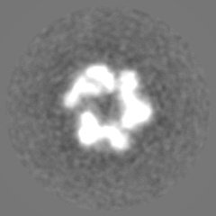
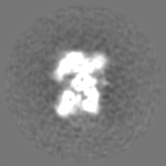
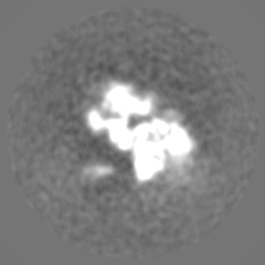
| 投影像・断面図 | 画像のコントロール
画像は Spider により作成 | ||||||||||||||||||||||||||||||||||||||||||||||||||||||||||||||||||||
| ボクセルのサイズ | X=Y=Z: 1.24 Å | ||||||||||||||||||||||||||||||||||||||||||||||||||||||||||||||||||||
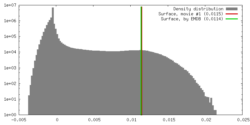
| 密度 |
| ||||||||||||||||||||||||||||||||||||||||||||||||||||||||||||||||||||
| 対称性 | 空間群: 1 | ||||||||||||||||||||||||||||||||||||||||||||||||||||||||||||||||||||
| 詳細 | EMDB XML:
CCP4マップ ヘッダ情報:
| ||||||||||||||||||||||||||||||||||||||||||||||||||||||||||||||||||||
 Z (Sec.)
 Y (Row.)
 X (Col.)


-添付データ
- 試料の構成要素
-全体 : Peroxisomal Pex1/Pex6 ATPase complex
| 全体 | 名称: Peroxisomal Pex1/Pex6 ATPase complex |
|---|---|
| 要素 |
|
-超分子 #1000: Peroxisomal Pex1/Pex6 ATPase complex
| 超分子 | 名称: Peroxisomal Pex1/Pex6 ATPase complex / タイプ: sample / ID: 1000 詳細: The sample was freshly prepared before being loaded onto grids. 集合状態: heterohexamer formed by three Pex1 and three Pex6 Number unique components: 2 |
|---|---|
| 分子量 | 理論値: 726 KDa |
-分子 #1: Peroxisomal ATPase Pex1
| 分子 | 名称: Peroxisomal ATPase Pex1 / タイプ: protein_or_peptide / ID: 1 / Name.synonym: Pex1 / コピー数: 3 集合状態: Three molecules of Pex1 interact with three molecules of Pex6 to form a heterohexamer of alternating subunits. 組換発現: Yes |
|---|---|
| 由来(天然) | 生物種: |
| 分子量 | 理論値: 122 KDa |
| 組換発現 | 生物種: |
| 配列 | UniProtKB: Peroxisomal ATPase PEX1 / InterPro: Peroxisome biogenesis factor 1 |
-分子 #2: Peroxisomal ATPase Pex6
| 分子 | 名称: Peroxisomal ATPase Pex6 / タイプ: protein_or_peptide / ID: 2 / Name.synonym: Pex6 / コピー数: 3 集合状態: Three molecules of Pex1 interact with three molecules of Pex6 to form a heterohexamer of alternating subunits. 組換発現: Yes |
|---|---|
| 由来(天然) | 生物種: |
| 分子量 | 理論値: 120 KDa |
| 組換発現 | 生物種: |
| 配列 | UniProtKB: Peroxisomal ATPase PEX6 |
-実験情報
-構造解析
| 手法 | クライオ電子顕微鏡法 |
|---|---|
 解析 解析 | 単粒子再構成法 |
| 試料の集合状態 | particle |
-試料調製
| 濃度 | 0.6 mg/mL |
|---|---|
| 緩衝液 | pH: 7.5 / 詳細: 40 mM Tris, pH 7.5, 25 mM NaCl, 4 mM ADP |
| グリッド | 詳細: Quantifoil 400 mesh holey carbon grid |
| 凍結 | 凍結剤: ETHANE / チャンバー内湿度: 90 % / チャンバー内温度: 90 K / 装置: FEI VITROBOT MARK I / 手法: Blot for 5 seconds before plunging |
- 電子顕微鏡法
| 顕微鏡 | FEI TECNAI F20 |
|---|---|
| 詳細 | Images were recorded using a Gatan K2 Summit in super-resolution counting mode. Motion correction as described in Li et al. (2013) Nature Methods. |
| 日付 | 2014年5月13日 |
| 撮影 | カテゴリ: CCD / フィルム・検出器のモデル: GATAN K2 (4k x 4k) / 実像数: 491 / 平均電子線量: 38 e/Å2 |
| 電子線 | 加速電圧: 200 kV / 電子線源:  FIELD EMISSION GUN FIELD EMISSION GUN |
| 電子光学系 | 倍率(補正後): 40410 / 照射モード: FLOOD BEAM / 撮影モード: BRIGHT FIELD / Cs: 2 mm / 最大 デフォーカス(公称値): -2.2 µm / 最小 デフォーカス(公称値): -0.9 µm / 倍率(公称値): 29000 |
| 試料ステージ | 試料ホルダー: Oxford instrument nitrogen-cooled side-entry holder 試料ホルダーモデル: GATAN LIQUID NITROGEN |
| 実験機器 |  モデル: Tecnai F20 / 画像提供: FEI Company |
-画像解析
| CTF補正 | 詳細: Each particle |
|---|---|
| 最終 再構成 | アルゴリズム: OTHER / 解像度のタイプ: BY AUTHOR / 解像度: 8.8 Å / 解像度の算出法: OTHER / ソフトウェア - 名称: SPARX, RELION 詳細: Initial model building was done in SPARX using the common-line method. 3D classification, refinement, and subsequent reconstruction were performed using RELION. 使用した粒子像数: 16117 |
