 Movie
Movie Controller

[English] 日本語
 Yorodumi
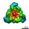
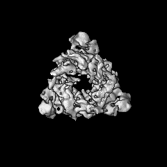
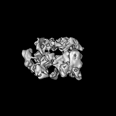
- EMDB-6360: Cryo-EM structure of the peroxisomal Pex1/Pex6 complex in ADP state -
+ Open data

- Basic information
| Entry | Database: EMDB / ID: EMD-6360 | |||||||||
|---|---|---|---|---|---|---|---|---|---|---|
| Title | Cryo-EM structure of the peroxisomal Pex1/Pex6 complex in ADP state | |||||||||
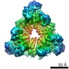
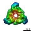
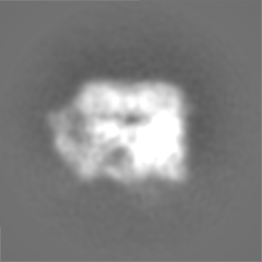
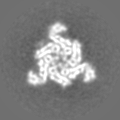
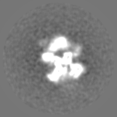
 Map data Map data | Reconstruction of Pex1/Pex6 AAA ATPase complex in ADP using single-particle cryo-electron microscopy without imposed symmetry | |||||||||
 Sample Sample |
| |||||||||
 Keywords Keywords | AAA ATPase / cryoelectron microscopy / peroxisome | |||||||||
| Function / homology |  Function and homology information Function and homology informationprotein import into peroxisome matrix, receptor recycling / protein import into peroxisome matrix / protein transporter activity / peroxisomal membrane / ATPase complex / protein unfolding / Hydrolases; Acting on acid anhydrides; Acting on acid anhydrides to facilitate cellular and subcellular movement / peroxisome / ATP hydrolysis activity / ATP binding / cytosol Similarity search - Function | |||||||||
| Biological species |  | |||||||||
| Method | single particle reconstruction / cryo EM / Resolution: 8.8 Å | |||||||||
 Authors Authors | Blok NB / Tan D / Wang RY / Penczek PA / Baker D / DiMaio F / Rapoport TA / Walz T | |||||||||
 Citation Citation | Journal: Proc Natl Acad Sci U S A / Year: 2015 Title: Unique double-ring structure of the peroxisomal Pex1/Pex6 ATPase complex revealed by cryo-electron microscopy. Authors: Neil B Blok / Dongyan Tan / Ray Yu-Ruei Wang / Pawel A Penczek / David Baker / Frank DiMaio / Tom A Rapoport / Thomas Walz /  Abstract: Members of the AAA family of ATPases assemble into hexameric double rings and perform vital functions, yet their molecular mechanisms remain poorly understood. Here, we report structures of the ...Members of the AAA family of ATPases assemble into hexameric double rings and perform vital functions, yet their molecular mechanisms remain poorly understood. Here, we report structures of the Pex1/Pex6 complex; mutations in these proteins frequently cause peroxisomal diseases. The structures were determined in the presence of different nucleotides by cryo-electron microscopy. Models were generated using a computational approach that combines Monte Carlo placement of structurally homologous domains into density maps with energy minimization and refinement protocols. Pex1 and Pex6 alternate in an unprecedented hexameric double ring. Each protein has two N-terminal domains, N1 and N2, structurally related to the single N domains in p97 and N-ethylmaleimide sensitive factor (NSF); N1 of Pex1 is mobile, but the others are packed against the double ring. The N-terminal ATPase domains are inactive, forming a symmetric D1 ring, whereas the C-terminal domains are active, likely in different nucleotide states, and form an asymmetric D2 ring. These results suggest how subunit activity is coordinated and indicate striking similarities between Pex1/Pex6 and p97, supporting the hypothesis that the Pex1/Pex6 complex has a role in peroxisomal protein import analogous to p97 in ER-associated protein degradation. | |||||||||
| History |
|
- Structure visualization
| Movie |
Movie viewer |
|---|---|
| Structure viewer | EM map: SurfViewMolmilJmol/JSmol |
| Supplemental images |

- Downloads & links
-EMDB archive
| Map data | emd_6360.map.gz | 40 MB | EMDB map data format | |
|---|---|---|---|---|
| Header (meta data) | emd-6360-v30.xmlemd-6360.xml | 11.8 KB 11.8 KB | Display Display | EMDB header |
| Images | 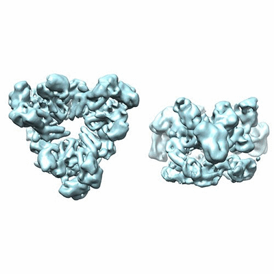 400_6360.gif 400_6360.gif 80_6360.gif 80_6360.gif | 49.9 KB 3.1 KB | ||
| Archive directory |  http://ftp.pdbj.org/pub/emdb/structures/EMD-6360ftp://ftp.pdbj.org/pub/emdb/structures/EMD-6360 http://ftp.pdbj.org/pub/emdb/structures/EMD-6360ftp://ftp.pdbj.org/pub/emdb/structures/EMD-6360 | HTTPS FTP |
-Validation report
| Summary document | emd_6360_validation.pdf.gz | 78.4 KB | Display | EMDB validaton report |
|---|---|---|---|---|
| Full document | emd_6360_full_validation.pdf.gz | 77.2 KB | Display | |
| Data in XML | emd_6360_validation.xml.gz | 494 B | Display | |
| Arichive directory | https://ftp.pdbj.org/pub/emdb/validation_reports/EMD-6360ftp://ftp.pdbj.org/pub/emdb/validation_reports/EMD-6360 | HTTPS FTP |
-Related structure data


-Links
| EMDB pages | EMDB (EBI/PDBe) / EMDataResource |
|---|---|
| Related items in Molecule of the Month |


-Map
| File | Download / File: emd_6360.map.gz / Format: CCP4 / Size: 51.5 MB / Type: IMAGE STORED AS FLOATING POINT NUMBER (4 BYTES) | ||||||||||||||||||||||||||||||||||||||||||||||||||||||||||||||||||||
|---|---|---|---|---|---|---|---|---|---|---|---|---|---|---|---|---|---|---|---|---|---|---|---|---|---|---|---|---|---|---|---|---|---|---|---|---|---|---|---|---|---|---|---|---|---|---|---|---|---|---|---|---|---|---|---|---|---|---|---|---|---|---|---|---|---|---|---|---|---|
| Annotation | Reconstruction of Pex1/Pex6 AAA ATPase complex in ADP using single-particle cryo-electron microscopy without imposed symmetry | ||||||||||||||||||||||||||||||||||||||||||||||||||||||||||||||||||||
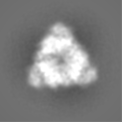
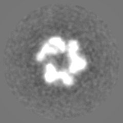
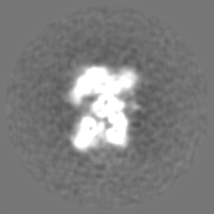
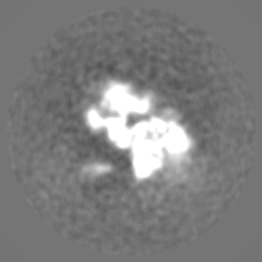
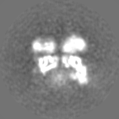
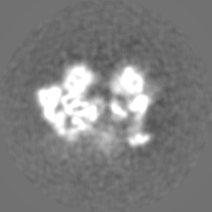
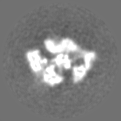
| Projections & slices | Image control
Images are generated by Spider. | ||||||||||||||||||||||||||||||||||||||||||||||||||||||||||||||||||||
| Voxel size | X=Y=Z: 1.24 Å | ||||||||||||||||||||||||||||||||||||||||||||||||||||||||||||||||||||
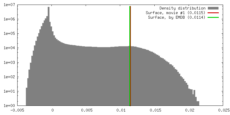
| Density |
| ||||||||||||||||||||||||||||||||||||||||||||||||||||||||||||||||||||
| Symmetry | Space group: 1 | ||||||||||||||||||||||||||||||||||||||||||||||||||||||||||||||||||||
| Details | EMDB XML:
CCP4 map header:
| ||||||||||||||||||||||||||||||||||||||||||||||||||||||||||||||||||||
 Z (Sec.)
 Y (Row.)
 X (Col.)


-Supplemental data
- Sample components
-Entire : Peroxisomal Pex1/Pex6 ATPase complex
| Entire | Name: Peroxisomal Pex1/Pex6 ATPase complex |
|---|---|
| Components |
|
-Supramolecule #1000: Peroxisomal Pex1/Pex6 ATPase complex
| Supramolecule | Name: Peroxisomal Pex1/Pex6 ATPase complex / type: sample / ID: 1000 Details: The sample was freshly prepared before being loaded onto grids. Oligomeric state: heterohexamer formed by three Pex1 and three Pex6 Number unique components: 2 |
|---|---|
| Molecular weight | Theoretical: 726 KDa |
-Macromolecule #1: Peroxisomal ATPase Pex1
| Macromolecule | Name: Peroxisomal ATPase Pex1 / type: protein_or_peptide / ID: 1 / Name.synonym: Pex1 / Number of copies: 3 Oligomeric state: Three molecules of Pex1 interact with three molecules of Pex6 to form a heterohexamer of alternating subunits. Recombinant expression: Yes |
|---|---|
| Source (natural) | Organism: |
| Molecular weight | Theoretical: 122 KDa |
| Recombinant expression | Organism: |
| Sequence | UniProtKB: Peroxisomal ATPase PEX1 / InterPro: Peroxisome biogenesis factor 1 |
-Macromolecule #2: Peroxisomal ATPase Pex6
| Macromolecule | Name: Peroxisomal ATPase Pex6 / type: protein_or_peptide / ID: 2 / Name.synonym: Pex6 / Number of copies: 3 Oligomeric state: Three molecules of Pex1 interact with three molecules of Pex6 to form a heterohexamer of alternating subunits. Recombinant expression: Yes |
|---|---|
| Source (natural) | Organism: |
| Molecular weight | Theoretical: 120 KDa |
| Recombinant expression | Organism: |
| Sequence | UniProtKB: Peroxisomal ATPase PEX6 |
-Experimental details
-Structure determination
| Method | cryo EM |
|---|---|
 Processing Processing | single particle reconstruction |
| Aggregation state | particle |
-Sample preparation
| Concentration | 0.6 mg/mL |
|---|---|
| Buffer | pH: 7.5 / Details: 40 mM Tris, pH 7.5, 25 mM NaCl, 4 mM ADP |
| Grid | Details: Quantifoil 400 mesh holey carbon grid |
| Vitrification | Cryogen name: ETHANE / Chamber humidity: 90 % / Chamber temperature: 90 K / Instrument: FEI VITROBOT MARK I / Method: Blot for 5 seconds before plunging |
- Electron microscopy
| Microscope | FEI TECNAI F20 |
|---|---|
| Details | Images were recorded using a Gatan K2 Summit in super-resolution counting mode. Motion correction as described in Li et al. (2013) Nature Methods. |
| Date | May 13, 2014 |
| Image recording | Category: CCD / Film or detector model: GATAN K2 (4k x 4k) / Number real images: 491 / Average electron dose: 38 e/Å2 |
| Electron beam | Acceleration voltage: 200 kV / Electron source:  FIELD EMISSION GUN FIELD EMISSION GUN |
| Electron optics | Calibrated magnification: 40410 / Illumination mode: FLOOD BEAM / Imaging mode: BRIGHT FIELD / Cs: 2 mm / Nominal defocus max: -2.2 µm / Nominal defocus min: -0.9 µm / Nominal magnification: 29000 |
| Sample stage | Specimen holder: Oxford instrument nitrogen-cooled side-entry holder Specimen holder model: GATAN LIQUID NITROGEN |
| Experimental equipment |  Model: Tecnai F20 / Image courtesy: FEI Company |
-Image processing
| CTF correction | Details: Each particle |
|---|---|
| Final reconstruction | Algorithm: OTHER / Resolution.type: BY AUTHOR / Resolution: 8.8 Å / Resolution method: OTHER / Software - Name: SPARX, RELION Details: Initial model building was done in SPARX using the common-line method. 3D classification, refinement, and subsequent reconstruction were performed using RELION. Number images used: 16117 |
