 ムービー
ムービー コントローラー


+ データを開く

- 基本情報
基本情報
| 登録情報 | データベース: EMDB / ID: EMD-6307 | |||||||||
|---|---|---|---|---|---|---|---|---|---|---|
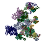
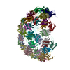
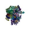
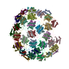
| タイトル | Electron cryo-microscopy of microtubule-bound TTLL7 | |||||||||
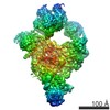
 マップデータ マップデータ | Reconstruction of a Tubulin Tyrosine Ligase-Like Glutamylase bound to the microtubule | |||||||||
 試料 試料 |
| |||||||||
 キーワード キーワード | microtubule-bound TTLL7 | |||||||||
| 生物種 |  Homo sapiens (ヒト) / Homo sapiens (ヒト) /  | |||||||||
| 手法 | らせん対称体再構成法 / クライオ電子顕微鏡法 / 解像度: 7.95 Å | |||||||||
 データ登録者 データ登録者 | Wilson-Kubalek EM / Garnham CP / Vemu A / Yu I / Szyk A / Lander GC / Milligan RA / Roll-Mecak A | |||||||||
 引用 引用 | ジャーナル: Cell / 年: 2015 タイトル: Multivalent Microtubule Recognition by Tubulin Tyrosine Ligase-like Family Glutamylases. 著者: Christopher P Garnham / Annapurna Vemu / Elizabeth M Wilson-Kubalek / Ian Yu / Agnieszka Szyk / Gabriel C Lander / Ronald A Milligan / Antonina Roll-Mecak /  要旨: Glutamylation, the most prevalent tubulin posttranslational modification, marks stable microtubules and regulates recruitment and activity of microtubule- interacting proteins. Nine enzymes of the ...Glutamylation, the most prevalent tubulin posttranslational modification, marks stable microtubules and regulates recruitment and activity of microtubule- interacting proteins. Nine enzymes of the tubulin tyrosine ligase-like (TTLL) family catalyze glutamylation. TTLL7, the most abundant neuronal glutamylase, adds glutamates preferentially to the β-tubulin tail. Coupled with ensemble and single-molecule biochemistry, our hybrid X-ray and cryo-electron microscopy structure of TTLL7 bound to the microtubule delineates a tripartite microtubule recognition strategy. The enzyme uses its core to engage the disordered anionic tails of α- and β-tubulin, and a flexible cationic domain to bind the microtubule and position itself for β-tail modification. Furthermore, we demonstrate that all single-chain TTLLs with known glutamylase activity utilize a cationic microtubule-binding domain analogous to that of TTLL7. Therefore, our work reveals the combined use of folded and intrinsically disordered substrate recognition elements as the molecular basis for specificity among the enzymes primarily responsible for chemically diversifying cellular microtubules. | |||||||||
| 履歴 |
|
- 構造の表示
構造の表示
| ムービー |
 ムービービューア ムービービューア |
|---|---|
| 構造ビューア | EMマップ: SurfViewMolmilJmol/JSmol |
| 添付画像 |

- ダウンロードとリンク
-EMDBアーカイブ
| マップデータ | emd_6307.map.gz | 1.5 MB | EMDBマップデータ形式 | |
|---|---|---|---|---|
| ヘッダ (付随情報) | emd-6307-v30.xmlemd-6307.xml | 10.7 KB 10.7 KB | 表示 表示 | EMDBヘッダ |
| 画像 |  400_6307.gif 400_6307.gif 80_6307.gif 80_6307.gif | 123.6 KB 7.1 KB | ||
| アーカイブディレクトリ |  http://ftp.pdbj.org/pub/emdb/structures/EMD-6307ftp://ftp.pdbj.org/pub/emdb/structures/EMD-6307 http://ftp.pdbj.org/pub/emdb/structures/EMD-6307ftp://ftp.pdbj.org/pub/emdb/structures/EMD-6307 | HTTPS FTP |
-検証レポート
| 文書・要旨 | emd_6307_validation.pdf.gz | 78.5 KB | 表示 | EMDB検証レポート |
|---|---|---|---|---|
| 文書・詳細版 | emd_6307_full_validation.pdf.gz | 77.6 KB | 表示 | |
| XML形式データ | emd_6307_validation.xml.gz | 494 B | 表示 | |
| アーカイブディレクトリ | https://ftp.pdbj.org/pub/emdb/validation_reports/EMD-6307ftp://ftp.pdbj.org/pub/emdb/validation_reports/EMD-6307 | HTTPS FTP |
-関連構造データ


-リンク
| EMDBのページ | EMDB (EBI/PDBe) / EMDataResource |
|---|
-マップ
| ファイル | ダウンロード / ファイル: emd_6307.map.gz / 形式: CCP4 / 大きさ: 1.6 MB / タイプ: IMAGE STORED AS FLOATING POINT NUMBER (4 BYTES) | ||||||||||||||||||||||||||||||||||||||||||||||||||||||||||||
|---|---|---|---|---|---|---|---|---|---|---|---|---|---|---|---|---|---|---|---|---|---|---|---|---|---|---|---|---|---|---|---|---|---|---|---|---|---|---|---|---|---|---|---|---|---|---|---|---|---|---|---|---|---|---|---|---|---|---|---|---|---|
| 注釈 | Reconstruction of a Tubulin Tyrosine Ligase-Like Glutamylase bound to the microtubule | ||||||||||||||||||||||||||||||||||||||||||||||||||||||||||||
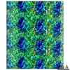
| 投影像・断面図 | 画像のコントロール
画像は Spider により作成 これらの図は立方格子座標系で作成されたものです | ||||||||||||||||||||||||||||||||||||||||||||||||||||||||||||
| ボクセルのサイズ | X=Y=Z: 2.73 Å | ||||||||||||||||||||||||||||||||||||||||||||||||||||||||||||
| 密度 |
| ||||||||||||||||||||||||||||||||||||||||||||||||||||||||||||
| 対称性 | 空間群: 1 | ||||||||||||||||||||||||||||||||||||||||||||||||||||||||||||
| 詳細 | EMDB XML:
CCP4マップ ヘッダ情報:
| ||||||||||||||||||||||||||||||||||||||||||||||||||||||||||||
 Z (Sec.)
 Y (Row.)
 X (Col.)


-添付データ
- 試料の構成要素
-全体 : Tubulin Tyrosine Ligase-Like (TTLL7) bound to the microtubule
| 全体 | 名称: Tubulin Tyrosine Ligase-Like (TTLL7) bound to the microtubule |
|---|---|
| 要素 |
|
-超分子 #1000: Tubulin Tyrosine Ligase-Like (TTLL7) bound to the microtubule
| 超分子 | 名称: Tubulin Tyrosine Ligase-Like (TTLL7) bound to the microtubule タイプ: sample / ID: 1000 / 詳細: The sample was monodisperse / 集合状態: one TTLL7 bound to tubulin dimer / Number unique components: 2 |
|---|---|
| 分子量 | 実験値: 61 KDa / 理論値: 61 KDa / 手法: SDS PAGE |
-分子 #1: Tubulin Tyrosine Ligase-Like (TTLL7) Family Glutamylase
| 分子 | 名称: Tubulin Tyrosine Ligase-Like (TTLL7) Family Glutamylase タイプ: protein_or_peptide / ID: 1 / Name.synonym: TTLL7 / コピー数: 1 / 集合状態: monomer / 組換発現: Yes |
|---|---|
| 由来(天然) | 生物種: Homo sapiens (ヒト) / 別称: Human |
| 分子量 | 実験値: 61 KDa / 理論値: 61 KDa |
| 組換発現 | 生物種:  |
-分子 #2: tubulin
| 分子 | 名称: tubulin / タイプ: protein_or_peptide / ID: 2 詳細: Two sources of tubulin were used: cytoskeletal tubulin from bovine brain and human tubulin from TCA 201 cells. 組換発現: No / データベース: NCBI |
|---|---|
| 由来(天然) | 生物種: |
-実験情報
-構造解析
| 手法 | クライオ電子顕微鏡法 |
|---|---|
 解析 解析 | らせん対称体再構成法 |
| 試料の集合状態 | particle |
-試料調製
| 濃度 | 11 mg/mL |
|---|---|
| 緩衝液 | pH: 7 / 詳細: 20 mM HEPES, 0.5 mM ATP, 1 mM TCEP, 50 mM NaCl |
| グリッド | 詳細: Protochips C-flat grid: holey carbon with 2 um holes and 400 mesh copper grid with 2 um spacing |
| 凍結 | 凍結剤: ETHANE / チャンバー内湿度: 80 % / チャンバー内温度: 93 K / 装置: HOMEMADE PLUNGER 手法: Blot grid from behind for 3 seconds before plunging. |
- 電子顕微鏡法
| 顕微鏡 | FEI TECNAI F20 |
|---|---|
| アライメント法 | Legacy - 非点収差: Objective lens astigmatism was corrected at 62000x magnification. |
| 日付 | 2014年8月5日 |
| 撮影 | カテゴリ: CCD フィルム・検出器のモデル: TVIPS TEMCAM-F416 (4k x 4k) デジタル化 - サンプリング間隔: 2.73 µm / 実像数: 614 / 平均電子線量: 20 e/Å2 / ビット/ピクセル: 16 |
| 電子線 | 加速電圧: 200 kV / 電子線源:  FIELD EMISSION GUN FIELD EMISSION GUN |
| 電子光学系 | 照射モード: FLOOD BEAM / 撮影モード: BRIGHT FIELD / 最大 デフォーカス(公称値): 2.5 µm / 最小 デフォーカス(公称値): 0.5 µm / 倍率(公称値): 62000 |
| 試料ステージ | 試料ホルダーモデル: GATAN LIQUID NITROGEN |
| 実験機器 |  モデル: Tecnai F20 / 画像提供: FEI Company |
-画像解析
| 詳細 | We used IHRSR adapted for microtubules with a dimer repeat. |
|---|---|
| 最終 再構成 | アルゴリズム: OTHER / 解像度のタイプ: BY AUTHOR / 解像度: 7.95 Å / 解像度の算出法: OTHER / ソフトウェア - 名称: EMAN2, FREALIGN 詳細: Final maps were calculated from three averaged data sets. |
| CTF補正 | 詳細: CTFIND v3 |
