[English] 日本語
Yorodumi
- PDB-4ylr: Tubulin Glutamylase -

+
Open data


ID or keywords:

Loading...

-
Basic information

Entry
Database: PDB / ID: 4ylr
TitleTubulin Glutamylase
ComponentsTubulin polyglutamylase TTLL7
KeywordsLIGASE / Enzyme
Function / homology
Function and homology information


tubulin-glutamic acid ligase activity / protein polyglutamylation / Ligases; Forming carbon-nitrogen bonds; Acid-amino-acid ligases (peptide synthases) / Carboxyterminal post-translational modifications of tubulin / beta-tubulin binding / alpha-tubulin binding / tubulin binding / cilium / microtubule cytoskeleton organization / nervous system development ...tubulin-glutamic acid ligase activity / protein polyglutamylation / Ligases; Forming carbon-nitrogen bonds; Acid-amino-acid ligases (peptide synthases) / Carboxyterminal post-translational modifications of tubulin / beta-tubulin binding / alpha-tubulin binding / tubulin binding / cilium / microtubule cytoskeleton organization / nervous system development / perikaryon / microtubule / cell differentiation / dendrite / ATP binding / metal ion binding / cytosol
Similarity search - Function
Tubulin-tyrosine ligase/Tubulin polyglutamylase / Tubulin-tyrosine ligase family / TTL domain profile.
Similarity search - Domain/homology
ADENOSINE-5'-DIPHOSPHATE / Tubulin polyglutamylase TTLL7
Similarity search - Component
Biological speciesHomo sapiens (human)
MethodX-RAY DIFFRACTION / SYNCHROTRON / MOLECULAR REPLACEMENT / Resolution: 2.55 Å
AuthorsGarnham, C.P. / Vemu, A. / Wilson-Kubalek, E.M. / Yu, I. / Szyk, A. / Lander, G.C. / Milligan, R.A. / Roll-Mecak, A.
Funding support United States, 1items
OrganizationGrant numberCountry
National Institutes of Health/National Institute of General Medical Sciences (NIH/NIGMS) United States
Citation
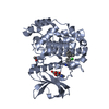
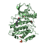
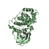
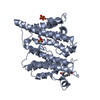
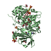
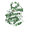
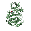
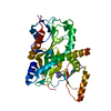
Journal: Cell / Year: 2015
Title: Multivalent Microtubule Recognition by Tubulin Tyrosine Ligase-like Family Glutamylases.
Authors: Christopher P Garnham / Annapurna Vemu / Elizabeth M Wilson-Kubalek / Ian Yu / Agnieszka Szyk / Gabriel C Lander / Ronald A Milligan / Antonina Roll-Mecak /
Abstract: Glutamylation, the most prevalent tubulin posttranslational modification, marks stable microtubules and regulates recruitment and activity of microtubule- interacting proteins. Nine enzymes of the ...Glutamylation, the most prevalent tubulin posttranslational modification, marks stable microtubules and regulates recruitment and activity of microtubule- interacting proteins. Nine enzymes of the tubulin tyrosine ligase-like (TTLL) family catalyze glutamylation. TTLL7, the most abundant neuronal glutamylase, adds glutamates preferentially to the β-tubulin tail. Coupled with ensemble and single-molecule biochemistry, our hybrid X-ray and cryo-electron microscopy structure of TTLL7 bound to the microtubule delineates a tripartite microtubule recognition strategy. The enzyme uses its core to engage the disordered anionic tails of α- and β-tubulin, and a flexible cationic domain to bind the microtubule and position itself for β-tail modification. Furthermore, we demonstrate that all single-chain TTLLs with known glutamylase activity utilize a cationic microtubule-binding domain analogous to that of TTLL7. Therefore, our work reveals the combined use of folded and intrinsically disordered substrate recognition elements as the molecular basis for specificity among the enzymes primarily responsible for chemically diversifying cellular microtubules.
History
DepositionMar 5, 2015Deposition site: RCSB / Processing site: RCSB
Revision 1.0Jun 17, 2015Provider: repository / Type: Initial release
Revision 1.1Sep 20, 2017Group: Author supporting evidence / Derived calculations / Source and taxonomy
Category: entity_src_gen / pdbx_audit_support / pdbx_struct_oper_list
Item: _entity_src_gen.pdbx_alt_source_flag / _pdbx_audit_support.funding_organization / _pdbx_struct_oper_list.symmetry_operation
Revision 1.2Dec 25, 2019Group: Author supporting evidence / Category: pdbx_audit_support / Item: _pdbx_audit_support.funding_organization
Revision 1.3Feb 28, 2024Group: Data collection / Database references / Category: chem_comp_atom / chem_comp_bond / database_2
Item: _database_2.pdbx_DOI / _database_2.pdbx_database_accession

-
Structure visualization

Structure viewerMolecule:
MolmilJmol/JSmol

Downloads & links

-
Assembly

Deposited unit
A: Tubulin polyglutamylase TTLL7
hetero molecules


Theoretical massNumber of molelcules
Total (without water)57,6942
Polymers57,2671
Non-polymers4271
Water27015
1


  • Idetical with deposited unit
  • defined by author&software
TypeNameSymmetry operationNumber
identity operation1_555x,y,z1
MethodPISA
Unit cell
Length a, b, c (Å)78.548, 121.762, 129.858
Angle α, β, γ (deg.)90.000, 90.000, 90.000
Int Tables number20
Space group name H-MC2221

-
Components

#1: Protein Tubulin polyglutamylase TTLL7 / Testis development protein NYD-SP30 / Tubulin--tyrosine ligase-like protein 7


Mass: 57266.715 Da / Num. of mol.: 1 / Fragment: UNP residues 36-518 / Mutation: E349Q
Source method: isolated from a genetically manipulated source
Source: (gene. exp.) Homo sapiens (human) / Gene: TTLL7 / Production host: Escherichia coli (E. coli) / References: UniProt: Q6ZT98, Ligases
#2: Chemical ChemComp-ADP / ADENOSINE-5'-DIPHOSPHATE


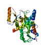
Mass: 427.201 Da / Num. of mol.: 1 / Source method: obtained synthetically / Formula: C10H15N5O10P2 / Comment: ADP, energy-carrying molecule*YM
#3: Water ChemComp-HOH / water


Mass: 18.015 Da / Num. of mol.: 15 / Source method: isolated from a natural source / Formula: H2O

-
Experimental details

-
Experiment

ExperimentMethod: X-RAY DIFFRACTION / Number of used crystals: 1

-
Sample preparation

CrystalDensity Matthews: 2.71 Å3/Da / Density % sol: 54.63 %
Crystal growTemperature: 289 K / Method: vapor diffusion, sitting drop / Details: PEG 8000, magnesium acetate

-
Data collection

DiffractionMean temperature: 100 K
Diffraction sourceSource: SYNCHROTRON / Site: ALS / Beamline: 5.0.1 / Wavelength: 0.9774 Å
DetectorType: ADSC QUANTUM 315r / Detector: CCD / Date: Dec 13, 2013
RadiationProtocol: SINGLE WAVELENGTH / Monochromatic (M) / Laue (L): M / Scattering type: x-ray
Radiation wavelengthWavelength: 0.9774 Å / Relative weight: 1
ReflectionResolution: 2.55→50 Å / Num. obs: 19556 / % possible obs: 93 % / Redundancy: 5.5 % / Biso Wilson estimate: 49.61 Å2 / Rmerge(I) obs: 0.124 / Rpim(I) all: 0.056 / Rrim(I) all: 0.137 / Χ2: 1.543 / Net I/av σ(I): 17.455 / Net I/σ(I): 7.8 / Num. measured all: 106539
Reflection shell

Diffraction-ID: 1 / Rejects: _

Resolution (Å)Redundancy (%)Rmerge(I) obsNum. unique allCC1/2Rpim(I) allRrim(I) allΧ2% possible all
2.55-2.593.90.4828150.8590.2560.5490.7579.4
2.59-2.644.20.4558440.9170.2350.5150.77382.3
2.64-2.694.30.4528950.9280.2310.5110.85186.7
2.69-2.754.70.4979120.9220.2410.5550.8290.4
2.75-2.814.90.4549830.930.2150.5040.86292.8
2.81-2.875.40.4219630.9480.1940.4660.91595.6
2.87-2.945.60.4229880.9510.190.4650.92496.1
2.94-3.025.70.35710100.9590.1590.3920.9696.1
3.02-3.115.90.2869770.9680.1280.3151.07997.2
3.11-3.215.90.26410010.9740.1180.2911.16596.6
3.21-3.335.90.21510070.9760.0960.2361.36296.1
3.33-3.4660.1889970.9810.0840.2071.55196
3.46-3.625.90.1589930.9830.0730.1751.80995.4
3.62-3.815.90.13810030.9790.0630.1522.02996.3
3.81-4.055.90.129990.9880.0540.1322.10995.7
4.05-4.365.80.1119910.9840.0510.1232.39894.7
4.36-4.85.90.1069950.9880.0470.1172.6494.8
4.8-5.495.90.1049910.9780.0480.1162.54494
5.49-6.925.70.08910100.9910.040.0981.88593.4
6.92-505.80.06610260.9940.0290.0731.90389.8

-
Processing

Software
NameVersionClassification
PHENIXrefinement
HKL-2000data reduction
SCALEPACKdata scaling
PHENIXphasing
PDB_EXTRACT3.15data extraction
RefinementMethod to determine structure: MOLECULAR REPLACEMENT / Resolution: 2.55→46.288 Å / SU ML: 0.31 / Cross valid method: FREE R-VALUE / σ(F): 1.36 / Phase error: 31.31 / Stereochemistry target values: ML
RfactorNum. reflection% reflection
Rfree0.2685 1980 10.12 %
Rwork0.2221 17576 -
obs0.2267 19556 94.56 %
Solvent computationShrinkage radii: 0.9 Å / VDW probe radii: 1.11 Å / Solvent model: FLAT BULK SOLVENT MODEL
Displacement parametersBiso max: 215.35 Å2 / Biso mean: 66.7442 Å2 / Biso min: 34.65 Å2
Refinement stepCycle: final / Resolution: 2.55→46.288 Å
ProteinNucleic acidLigandSolventTotal
Num. atoms2557 0 27 15 2599
Biso mean--105.42 55.29 -
Num. residues----331
Refine LS restraints
Refine-IDTypeDev idealNumber
X-RAY DIFFRACTIONf_bond_d0.0022653
X-RAY DIFFRACTIONf_angle_d0.5633609
X-RAY DIFFRACTIONf_chiral_restr0.021407
X-RAY DIFFRACTIONf_plane_restr0.002456
X-RAY DIFFRACTIONf_dihedral_angle_d13.281933
LS refinement shell

Refine-ID: X-RAY DIFFRACTION / Total num. of bins used: 14

Resolution (Å)Rfactor RfreeNum. reflection RfreeRfactor RworkNum. reflection RworkNum. reflection all% reflection obs (%)
2.55-2.61380.32491420.26491109125185
2.6138-2.68440.3131340.25721153128789
2.6844-2.76340.3281220.24991243136594
2.7634-2.85260.29791440.25251251139597
2.8526-2.95450.31341570.24111269142697
2.9545-3.07280.28691500.23821261141197
3.0728-3.21260.28851610.2281263142497
3.2126-3.3820.28011470.221277142497
3.382-3.59380.25911250.23641293141896
3.5938-3.87110.25331340.21121275140996
3.8711-4.26040.25081420.20881291143396
4.2604-4.87640.2351440.19241278142295
4.8764-6.14140.2731460.22641289143595
6.1414-46.29530.25751320.22081324145692
Refinement TLS params.

Method: refined / Refine-ID: X-RAY DIFFRACTION

IDL112)L122)L132)L222)L232)L332)S11 (Å °)S12 (Å °)S13 (Å °)S21 (Å °)S22 (Å °)S23 (Å °)S31 (Å °)S32 (Å °)S33 (Å °)T112)T122)T132)T222)T232)T332)Origin x (Å)Origin y (Å)Origin z (Å)
14.02182.2164-4.62084.6745-2.15485.4327-0.19341.3285-0.2123-0.24630.35680.2310.1053-1.207-0.12960.4509-0.0027-0.08610.859-0.14360.4735-29.3935-17.0813-38.6734
25.0566-0.87661.08552.11640.58655.04650.08050.1721-0.5338-0.06060.0250.03610.34940.1331-0.12050.3436-0.07460.00830.5313-0.01480.4544-16.8303-19.8912-27.6651
33.276-2.1260.7884.7-1.3130.4115-0.058-0.6858-0.71880.5428-0.8176-0.7590.7543-0.31990.68510.9015-0.07170.06191.12280.36850.8694-16.828-30.42681.8682
45.6075-4.6913-2.17837.48851.68512.7646-0.1061-0.8581-0.90140.7239-0.07391.1230.2963-0.37410.15090.4982-0.0367-0.03120.92620.08320.5971-26.3647-22.7316-2.6895
53.94680.3871-3.63741.67350.05636.3261-0.0029-0.53380.11630.1872-0.15220.1795-0.0276-0.29290.19060.3809-0.0224-0.03540.6324-0.10640.3504-24.3851-11.3572-17.1662
66.3354-2.6772-2.98821.36171.82494.30960.0416-0.27280.02970.0338-0.0126-0.0654-0.1285-0.1488-0.01570.426-0.0549-0.02020.5306-0.00720.4175-18.2637-11.5677-18.3706
75.2275-2.42392.23868.95782.27194.1575-0.20450.5667-0.11410.25460.0729-0.1322-0.0197-0.75120.04580.3644-0.0075-0.03940.53030.0140.3178-28.9158-9.2977-30.5398
89.49685.06793.17573.24153.58168.56270.2856-0.86331.5189-0.1313-0.1476-0.1396-1.05970.29310.04190.5457-0.15890.09260.5819-0.0670.5922-11.6107-2.1904-20.8763
98.845-4.1209-0.57139.74072.12529.71370.2296-1.47590.09350.35630.0652-1.60560.08640.6342-0.29090.3117-0.0595-0.02580.82210.13020.5519-0.8762-17.5241-17.5393
Refinement TLS group
IDRefine-IDRefine TLS-IDSelection detailsAuth asym-IDAuth seq-ID
1X-RAY DIFFRACTION1chain 'A' and (resid 40 through 54 )A0
2X-RAY DIFFRACTION2chain 'A' and (resid 55 through 133 )A0
3X-RAY DIFFRACTION3chain 'A' and (resid 134 through 161 )A0
4X-RAY DIFFRACTION4chain 'A' and (resid 169 through 200 )A0
5X-RAY DIFFRACTION5chain 'A' and (resid 201 through 235 )A0
6X-RAY DIFFRACTION6chain 'A' and (resid 269 through 352 )A0
7X-RAY DIFFRACTION7chain 'A' and (resid 353 through 382 )A0
8X-RAY DIFFRACTION8chain 'A' and (resid 451 through 468 )A0
9X-RAY DIFFRACTION9chain 'A' and (resid 469 through 484 )A0

+
About Yorodumi

-
News

-
Feb 9, 2022. New format data for meta-information of EMDB entries

New format data for meta-information of EMDB entries

  • Version 3 of the EMDB header file is now the official format.
  • The previous official version 1.9 will be removed from the archive.

Related info.:EMDB header

External links:wwPDB to switch to version 3 of the EMDB data model

-
Aug 12, 2020. Covid-19 info

Covid-19 info

URL: https://pdbjlvh1.pdbj.org/emnavi/covid19.php

New page: Covid-19 featured information page in EM Navigator.

Related info.:Covid-19 info / Mar 5, 2020. Novel coronavirus structure data

+
Mar 5, 2020. Novel coronavirus structure data

Novel coronavirus structure data

Related info.:Yorodumi Speices / Aug 12, 2020. Covid-19 info

External links:COVID-19 featured content - PDBj / Molecule of the Month (242):Coronavirus Proteases

+
Jan 31, 2019. EMDB accession codes are about to change! (news from PDBe EMDB page)

EMDB accession codes are about to change! (news from PDBe EMDB page)

  • The allocation of 4 digits for EMDB accession codes will soon come to an end. Whilst these codes will remain in use, new EMDB accession codes will include an additional digit and will expand incrementally as the available range of codes is exhausted. The current 4-digit format prefixed with “EMD-” (i.e. EMD-XXXX) will advance to a 5-digit format (i.e. EMD-XXXXX), and so on. It is currently estimated that the 4-digit codes will be depleted around Spring 2019, at which point the 5-digit format will come into force.
  • The EM Navigator/Yorodumi systems omit the EMD- prefix.

Related info.:Q: What is EMD? / ID/Accession-code notation in Yorodumi/EM Navigator

External links:EMDB Accession Codes are Changing Soon! / Contact to PDBj

+
Jul 12, 2017. Major update of PDB

Major update of PDB

  • wwPDB released updated PDB data conforming to the new PDBx/mmCIF dictionary.
  • This is a major update changing the version number from 4 to 5, and with Remediation, in which all the entries are updated.
  • In this update, many items about electron microscopy experimental information are reorganized (e.g. em_software).
  • Now, EM Navigator and Yorodumi are based on the updated data.

External links:wwPDB Remediation / Enriched Model Files Conforming to OneDep Data Standards Now Available in the PDB FTP Archive

-
Yorodumi

Thousand views of thousand structures

  • Yorodumi is a browser for structure data from EMDB, PDB, SASBDB, etc.
  • This page is also the successor to EM Navigator detail page, and also detail information page/front-end page for Omokage search.
  • The word "yorodu" (or yorozu) is an old Japanese word meaning "ten thousand". "mi" (miru) is to see.

Related info.:EMDB / PDB / SASBDB / Comparison of 3 databanks / Yorodumi Search / Aug 31, 2016. New EM Navigator & Yorodumi / Yorodumi Papers / Jmol/JSmol / Function and homology information / Changes in new EM Navigator and Yorodumi

Read more