 Movie
Movie Controller


+ Open data

- Basic information
| Entry |  | |||||||||
|---|---|---|---|---|---|---|---|---|---|---|
| Title | Cryo-EM structure of bacteriophage phiXacJX1 capsid | |||||||||
 Map data Map data | ||||||||||
 Sample Sample |
| |||||||||
 Keywords Keywords | Xanthomonas phage / capsid / icosahedron / VIRUS | |||||||||
| Biological species |  Xanthomonas phage phiXacJX1 (virus) Xanthomonas phage phiXacJX1 (virus) | |||||||||
| Method | single particle reconstruction / cryo EM / Resolution: 3.5 Å | |||||||||
 Authors Authors | Guo M / Wang A / Zheng Y / Liu C / Shao Q / Fang Q | |||||||||
| Funding support | 1 items
| |||||||||
 Citation Citation | Journal: Structure / Year: 2025 Title: Cryo-EM structures of a Xanthomonas phage: Insights into viral architecture and implications for the model phage HK97. Authors: Mingcheng Guo / Aohan Wang / Yaqi Zheng / Chaoying Liu / Qianqian Shao / Yunfei Deng / Lin Li / Yueting Wang / Xiaofang Wang / Yue Shen / Jun Qian / Xiaofeng Zhou / Qianglin Fang /  Abstract: Xanthomonas bacteria are responsible for disease outbreaks in several hundred plant species, causing significant economic losses. Xanthomonas phages have emerged as a promising biocontrol strategy in ...Xanthomonas bacteria are responsible for disease outbreaks in several hundred plant species, causing significant economic losses. Xanthomonas phages have emerged as a promising biocontrol strategy in managing various important plant diseases caused by Xanthomonas bacteria. However, structural information for Xanthomonas phages has remained limited so far. Here, we present high-resolution cryo-electron microscopy (cryo-EM) structures of the Xanthomonas citri phage ΦXacJX1 from siphoviruses. These structures include atomic models for the head, head-to-tail connector and head-proximal portion of the tail. ΦXacJX1's head and head-to-tail connector components show significant protein sequence and structural homology with those of the model siphophage HK97. However, the in-situ structures of head-to-tail connector of phage HK97 remain unavailable. The presented structures of phage ΦXacJX1 enhance our understanding of Xanthomonas phages and the mature virion of phage HK97. They provide a valuable framework for future structural and functional studies on both Xanthomonas phages and phage HK97. | |||||||||
| History |
|
- Structure visualization
| Supplemental images |
|---|

- Downloads & links
-EMDB archive
| Map data | emd_62947.map.gz | 1.1 GB |  EMDB map data format EMDB map data format | |
|---|---|---|---|---|
| Header (meta data) | emd-62947-v30.xmlemd-62947.xml | 16.2 KB 16.2 KB | Display Display | EMDB header |
| FSC (resolution estimation) | emd_62947_fsc.xml | 23.6 KB | Display | FSC data file |
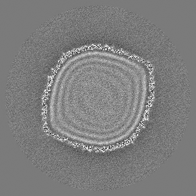
| Images |  emd_62947.png emd_62947.png | 154.1 KB | ||
| Masks | emd_62947_msk_1.map | 1.1 GB | Mask map | |
| Filedesc metadata | emd-62947.cif.gz | 5.9 KB | ||
| Others | emd_62947_half_map_1.map.gzemd_62947_half_map_2.map.gz | 942.3 MB 945.8 MB | ||
| Archive directory |  http://ftp.pdbj.org/pub/emdb/structures/EMD-62947ftp://ftp.pdbj.org/pub/emdb/structures/EMD-62947 http://ftp.pdbj.org/pub/emdb/structures/EMD-62947ftp://ftp.pdbj.org/pub/emdb/structures/EMD-62947 | HTTPS FTP |
-Related structure data


-Links
| EMDB pages | EMDB (EBI/PDBe) / EMDataResource |
|---|
-Map
| File | Download / File: emd_62947.map.gz / Format: CCP4 / Size: 1.1 GB / Type: IMAGE STORED AS FLOATING POINT NUMBER (4 BYTES) | ||||||||||||||||||||||||||||||||||||
|---|---|---|---|---|---|---|---|---|---|---|---|---|---|---|---|---|---|---|---|---|---|---|---|---|---|---|---|---|---|---|---|---|---|---|---|---|---|
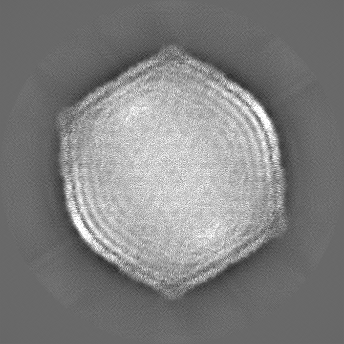
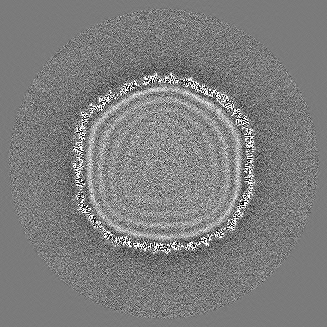
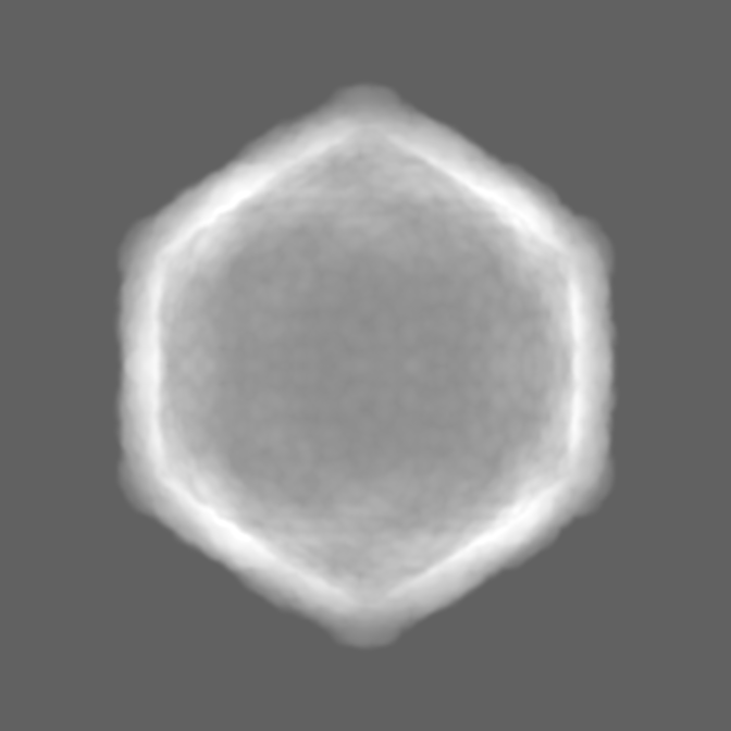
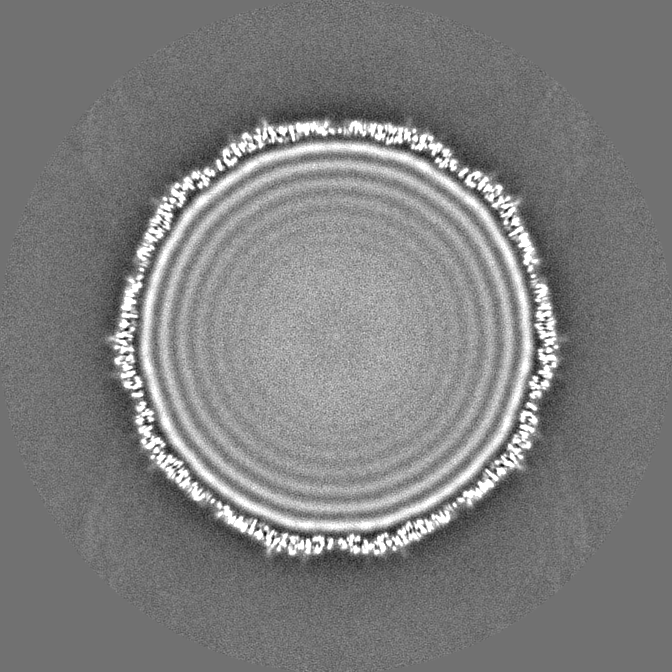
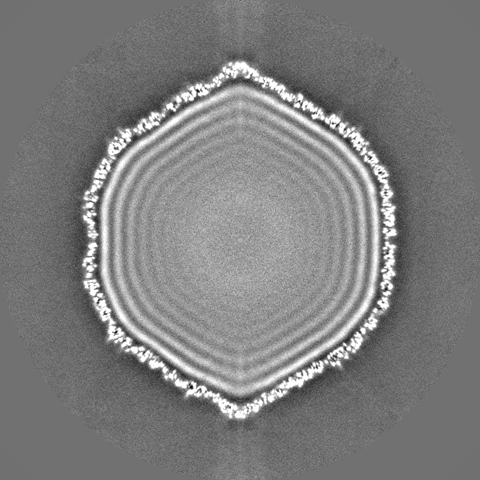
| Projections & slices | Image control
Images are generated by Spider. | ||||||||||||||||||||||||||||||||||||
| Voxel size | X=Y=Z: 1.372 Å | ||||||||||||||||||||||||||||||||||||
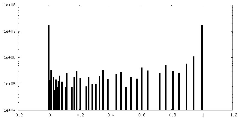
| Density |
| ||||||||||||||||||||||||||||||||||||
| Symmetry | Space group: 1 | ||||||||||||||||||||||||||||||||||||
| Details | EMDB XML:
|
 Z (Sec.)
 Y (Row.)
 X (Col.)


-Supplemental data
-Mask #1
| File | emd_62947_msk_1.map | ||||||||||||
|---|---|---|---|---|---|---|---|---|---|---|---|---|---|
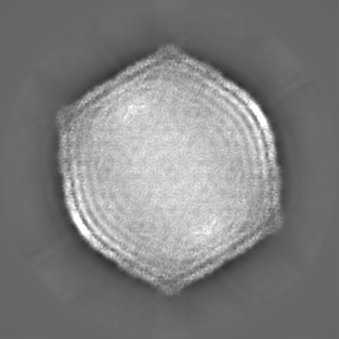
| Projections & Slices |
| ||||||||||||
| Density Histograms |


-Half map: #2
| File | emd_62947_half_map_1.map | ||||||||||||
|---|---|---|---|---|---|---|---|---|---|---|---|---|---|
| Projections & Slices |
| ||||||||||||
| Density Histograms |


-Half map: #1
| File | emd_62947_half_map_2.map | ||||||||||||
|---|---|---|---|---|---|---|---|---|---|---|---|---|---|
| Projections & Slices |
| ||||||||||||
| Density Histograms |


- Sample components
-Entire : Xanthomonas phage phiXacJX1
| Entire | Name: Xanthomonas phage phiXacJX1 (virus) |
|---|---|
| Components |
|
-Supramolecule #1: Xanthomonas phage phiXacJX1
| Supramolecule | Name: Xanthomonas phage phiXacJX1 / type: virus / ID: 1 / Parent: 0 / Macromolecule list: all / NCBI-ID: 3374911 / Sci species name: Xanthomonas phage phiXacJX1 / Virus type: VIRION / Virus isolate: STRAIN / Virus enveloped: No / Virus empty: No |
|---|---|
| Host (natural) | Organism:  Xanthomonas citri pv. citri (bacteria) Xanthomonas citri pv. citri (bacteria) |
-Macromolecule #1: major capsid protein gp3
| Macromolecule | Name: major capsid protein gp3 / type: protein_or_peptide / ID: 1 Details: This protein corresponds to a novel sequence that is not yet available in UniProt. The current reference for the sequence is GenBank Accession Number: PQ476032.1 Number of copies: 7 / Enantiomer: LEVO |
|---|---|
| Source (natural) | Organism: Xanthomonas phage phiXacJX1 (virus) |
| Molecular weight | Theoretical: 41.850199 KDa |
| Sequence | String: MSDINVINST LANISDSLKA HADRAVKEQE LNASVRAKVD ELLMAQGALQ ADLKAAQQRI AEVEGNGAGG DVQHISIGQQ FVNSDSFKA MAESGGQRGR AELNIKAAIT SLTTNADGSA GATVQTTRLP GIRELPQRRM TIRSLLAQGT MEGNTLEYVR E TGFTNAAA ...String: MSDINVINST LANISDSLKA HADRAVKEQE LNASVRAKVD ELLMAQGALQ ADLKAAQQRI AEVEGNGAGG DVQHISIGQQ FVNSDSFKA MAESGGQRGR AELNIKAAIT SLTTNADGSA GATVQTTRLP GIRELPQRRM TIRSLLAQGT MEGNTLEYVR E TGFTNAAA PVAEGAQKPE SSLKFDLVQT SAKVIAHWMK ASRQILSDSA QLQSFINARL LRGLEVVEEN QLLNGNGTGQ NL LGLLPQA TAFAAPITVA NATAVDRLRL ALLQAQLAEF PATGIVLNPA DWAGIELLKD TQGRYILGNP QGTLAPTLWG LPV VATQAM AAGQFLTGAF DAGAQVFDRW AARVEVATEN QDDFIKNMVT ILAEERLALA VYRPESFIKG SLTAAAAT |
-Experimental details
-Structure determination
| Method | cryo EM |
|---|---|
 Processing Processing | single particle reconstruction |
| Aggregation state | particle |
-Sample preparation
| Buffer | pH: 7.5 |
|---|---|
| Grid | Model: Quantifoil R2/1 / Material: COPPER / Support film - Material: CARBON / Support film - topology: CONTINUOUS / Pretreatment - Type: GLOW DISCHARGE |
| Vitrification | Cryogen name: ETHANE / Instrument: FEI VITROBOT MARK IV |
- Electron microscopy
| Microscope | TFS KRIOS |
|---|---|
| Image recording | Film or detector model: FEI FALCON IV (4k x 4k) / Average electron dose: 26.0 e/Å2 |
| Electron beam | Acceleration voltage: 300 kV / Electron source:  FIELD EMISSION GUN FIELD EMISSION GUN |
| Electron optics | Illumination mode: FLOOD BEAM / Imaging mode: BRIGHT FIELD / Nominal defocus max: 3.0 µm / Nominal defocus min: 1.0 µm / Nominal magnification: 59000 |
| Sample stage | Specimen holder model: FEI TITAN KRIOS AUTOGRID HOLDER |
| Experimental equipment |  Model: Titan Krios / Image courtesy: FEI Company |

