 Movie
Movie Controller

[English] 日本語
 Yorodumi
- EMDB-62860: CryoEM structure of class Ib Ribonucleotide reductase from Mycoba... -
+ Open data

- Basic information
| Entry |  | |||||||||
|---|---|---|---|---|---|---|---|---|---|---|
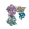
| Title | CryoEM structure of class Ib Ribonucleotide reductase from Mycobacterium thermoresistibile, conformer D | |||||||||
 Map data Map data | ||||||||||
 Sample Sample |
| |||||||||
 Keywords Keywords | Enzyme complex / supply Dexoyribonucleotides for replication and repair / asymmetric complex / REPLICATION | |||||||||
| Biological species |  Mycolicibacterium thermoresistibile ATCC 19527 (bacteria) Mycolicibacterium thermoresistibile ATCC 19527 (bacteria) | |||||||||
| Method | single particle reconstruction / cryo EM / Resolution: 3.8 Å | |||||||||
 Authors Authors | Yadav LR / Mande SC / Vinothkumar KR / Kumar J | |||||||||
| Funding support |  India, 2 items India, 2 items
| |||||||||
 Citation Citation | Journal: To Be Published Title: Dynamics of Class Ib ribonucleotide reductase complex revealed by CryoEM Authors: Yadav LR / Mande SC / Vinothkumar KR / Kumar J | |||||||||
| History |
|
- Structure visualization
| Supplemental images |
|---|

- Downloads & links
-EMDB archive
| Map data | emd_62860.map.gz | 86 MB |  EMDB map data format EMDB map data format | |
|---|---|---|---|---|
| Header (meta data) | emd-62860-v30.xmlemd-62860.xml | 24.4 KB 24.4 KB | Display Display | EMDB header |
| FSC (resolution estimation) | emd_62860_fsc.xml | 9.5 KB | Display | FSC data file |
| Images |  emd_62860.png emd_62860.png | 176.8 KB | ||
| Filedesc metadata | emd-62860.cif.gz | 7.2 KB | ||
| Others | emd_62860_additional_1.map.gzemd_62860_half_map_1.map.gzemd_62860_half_map_2.map.gz | 45.4 MB 84.7 MB 84.7 MB | ||
| Archive directory |  http://ftp.pdbj.org/pub/emdb/structures/EMD-62860ftp://ftp.pdbj.org/pub/emdb/structures/EMD-62860 http://ftp.pdbj.org/pub/emdb/structures/EMD-62860ftp://ftp.pdbj.org/pub/emdb/structures/EMD-62860 | HTTPS FTP |
-Related structure data
| Related structure data |  9l6oM M: atomic model generated by this map |
|---|
-Links
| EMDB pages | EMDB (EBI/PDBe) / EMDataResource |
|---|
-Map
| File | Download / File: emd_62860.map.gz / Format: CCP4 / Size: 91.1 MB / Type: IMAGE STORED AS FLOATING POINT NUMBER (4 BYTES) | ||||||||||||||||||||||||||||||||||||
|---|---|---|---|---|---|---|---|---|---|---|---|---|---|---|---|---|---|---|---|---|---|---|---|---|---|---|---|---|---|---|---|---|---|---|---|---|---|
| Projections & slices | Image control
Images are generated by Spider. | ||||||||||||||||||||||||||||||||||||
| Voxel size | X=Y=Z: 1.065 Å | ||||||||||||||||||||||||||||||||||||
| Density |
| ||||||||||||||||||||||||||||||||||||
| Symmetry | Space group: 1 | ||||||||||||||||||||||||||||||||||||
| Details | EMDB XML:
|
 Z (Sec.)
 Y (Row.)
 X (Col.)


-Supplemental data
-Additional map: #1
| File | emd_62860_additional_1.map | ||||||||||||
|---|---|---|---|---|---|---|---|---|---|---|---|---|---|
| Projections & Slices |
| ||||||||||||
| Density Histograms |


-Half map: #2
| File | emd_62860_half_map_1.map | ||||||||||||
|---|---|---|---|---|---|---|---|---|---|---|---|---|---|
| Projections & Slices |
| ||||||||||||
| Density Histograms |


-Half map: #1
| File | emd_62860_half_map_2.map | ||||||||||||
|---|---|---|---|---|---|---|---|---|---|---|---|---|---|
| Projections & Slices |
| ||||||||||||
| Density Histograms |


- Sample components
-Entire : Binary complex of RNR
| Entire | Name: Binary complex of RNR |
|---|---|
| Components |
|
-Supramolecule #1: Binary complex of RNR
| Supramolecule | Name: Binary complex of RNR / type: complex / ID: 1 / Parent: 0 / Macromolecule list: #1-#2 / Details: Heterotetrameric asymmetric complex of RNR |
|---|---|
| Source (natural) | Organism: Mycolicibacterium thermoresistibile ATCC 19527 (bacteria) |
-Macromolecule #1: Ribonucleoside-diphosphate reductase subunit beta
| Macromolecule | Name: Ribonucleoside-diphosphate reductase subunit beta / type: protein_or_peptide / ID: 1 / Number of copies: 2 / Enantiomer: LEVO / EC number: ribonucleoside-diphosphate reductase |
|---|---|
| Source (natural) | Organism: Mycolicibacterium thermoresistibile ATCC 19527 (bacteria) |
| Molecular weight | Theoretical: 36.487891 KDa |
| Recombinant expression | Organism: |
| Sequence | String: MKLVNRVSAI NWNRLEDDKD AEVWHRLTGN FWLPEKVPVS NDIPSWNTLT DTEKQLTMRV FTGLTLLDTI QGTVGAVSLI PDALTPHEE AVLTNIAFME SVHAKSYSNI FSTLCSTAEI DDAFRWSEEN PNLQRKAEIV MQYYRGDEPL KRKVASTLLE S FLFYSGFY ...String: MKLVNRVSAI NWNRLEDDKD AEVWHRLTGN FWLPEKVPVS NDIPSWNTLT DTEKQLTMRV FTGLTLLDTI QGTVGAVSLI PDALTPHEE AVLTNIAFME SVHAKSYSNI FSTLCSTAEI DDAFRWSEEN PNLQRKAEIV MQYYRGDEPL KRKVASTLLE S FLFYSGFY LPMYWSSRAK LTNTADMIRL IIRDEAVHGY YIGYKYQRGL AMVEPAKQKE LKDYTYDLLF ELYDNEVEYT QD LYDDVGL TEDVKKFLRY NANKALMNLG YEALFPKDET DVNPAILSAL APNADENHDF FSGSGSSYVI GKAVVTEDED W UniProtKB: Ribonucleoside-diphosphate reductase subunit beta |
-Macromolecule #2: Ribonucleoside-diphosphate reductase
| Macromolecule | Name: Ribonucleoside-diphosphate reductase / type: protein_or_peptide / ID: 2 / Number of copies: 2 / Enantiomer: LEVO / EC number: ribonucleoside-diphosphate reductase |
|---|---|
| Source (natural) | Organism: Mycolicibacterium thermoresistibile ATCC 19527 (bacteria) |
| Molecular weight | Theoretical: 76.554211 KDa |
| Recombinant expression | Organism: |
| Sequence | String: DGKIQFDKDK QAAREFFLQH VNQNTVFFHD YDEKLDYLIE NDYYEPEVLD QYSRDFVKSL LDRAYAKKFR FPTFLGAFKY YTSYTLKTF DGKRYLERFE DRVVMVALTL AAGDVELAEK LVDEIMDGRF QPATPTFLNS GKKQRGEPVS CFLLRIEDNM E SIGRAINS ...String: DGKIQFDKDK QAAREFFLQH VNQNTVFFHD YDEKLDYLIE NDYYEPEVLD QYSRDFVKSL LDRAYAKKFR FPTFLGAFKY YTSYTLKTF DGKRYLERFE DRVVMVALTL AAGDVELAEK LVDEIMDGRF QPATPTFLNS GKKQRGEPVS CFLLRIEDNM E SIGRAINS ALQLSKRGGG VALLLSNVRE FGAPIKNIEN QSSGVIPIMK LLEDSFSYAN QLGARQGAGA VYLHAHHPDI YR FLDTKRE NADEKIRIKT LSLGVVIPDI TFELAKKNED MYLFSPYDVE RVYGVPFADI SVTEKYYEMV DNPRIRKSKI NAR EFFQTL AELQFESGYP YIMFEDTVNR SNPIEGKVTH SNLCSEILQV STPSEFNDDL SYKVVGKDIS CNLGSLNIAK AMDS PDFGQ TVEVAIRALT AVSDQTRIDS VPSIVRGNDE SHSIGLGQMN LHGYLGRERI FYGSEEAIDF TNMYFYTVCY HAVRA SNRI AIERGKHFVG FEKSKYATGE FFDKYTDQVW EPKTDKVREL FAKANIHIPT QEDWRRLKES VQKHGIYNAY LQAVPP TGS ISYINHSTSS IHPIASKIEI RKEGKIGRVY YPAPYMTNDN LEYFQDAYEI GYEKIIDTYA AATQHVDQGL SLTLFFK DT ATTRDVNKAQ IYAWRKGIKT LYYIR UniProtKB: Ribonucleoside-diphosphate reductase |
-Macromolecule #3: MANGANESE (II) ION
| Macromolecule | Name: MANGANESE (II) ION / type: ligand / ID: 3 / Number of copies: 4 / Formula: MN |
|---|---|
| Molecular weight | Theoretical: 54.938 Da |
-Experimental details
-Structure determination
| Method | cryo EM |
|---|---|
 Processing Processing | single particle reconstruction |
| Aggregation state | particle |
-Sample preparation
| Concentration | 1.25 mg/mL |
|---|---|
| Buffer | pH: 8 / Details: 25mM Tris pH8, 150mM NaCl |
| Grid | Model: Quantifoil R0.6/1 / Material: GOLD / Mesh: 300 / Support film - Material: CARBON / Support film - topology: HOLEY / Pretreatment - Type: GLOW DISCHARGE / Pretreatment - Time: 90 sec. / Pretreatment - Atmosphere: AIR |
| Vitrification | Cryogen name: ETHANE / Chamber humidity: 100 % / Chamber temperature: 291 K / Instrument: FEI VITROBOT MARK IV |
| Details | sampled crosslinked with glutaraldehyde |
- Electron microscopy
| Microscope | TFS KRIOS |
|---|---|
| Specialist optics | Phase plate: OTHER / Energy filter - Name: GIF Bioquantum / Energy filter - Slit width: 20 eV |
| Image recording | Film or detector model: GATAN K2 QUANTUM (4k x 4k) / Detector mode: COUNTING / Digitization - Dimensions - Width: 3710 pixel / Digitization - Dimensions - Height: 3838 pixel / Number grids imaged: 1 / Number real images: 831 / Average exposure time: 8.0 sec. / Average electron dose: 47.36 e/Å2 |
| Electron beam | Acceleration voltage: 300 kV / Electron source:  FIELD EMISSION GUN FIELD EMISSION GUN |
| Electron optics | C2 aperture diameter: 50.0 µm / Illumination mode: SPOT SCAN / Imaging mode: BRIGHT FIELD / Cs: 2.7 mm / Nominal defocus max: 3.5 µm / Nominal defocus min: 1.5 µm |
| Sample stage | Specimen holder model: FEI TITAN KRIOS AUTOGRID HOLDER / Cooling holder cryogen: NITROGEN |
| Experimental equipment |  Model: Titan Krios / Image courtesy: FEI Company |
+Image processing

-Atomic model buiding 1
| Initial model |
| ||||||
|---|---|---|---|---|---|---|---|
| Refinement | Space: REAL / Protocol: FLEXIBLE FIT | ||||||
| Output model | PDB-9l6o: |

