 Movie
Movie Controller


+ Open data

- Basic information
| Entry |  | |||||||||
|---|---|---|---|---|---|---|---|---|---|---|
| Title | Cryo-EM strucuture of human OAT1 in complex with probenecid | |||||||||
 Map data Map data | ||||||||||
 Sample Sample |
| |||||||||
 Keywords Keywords | membrane transporter / antiporter / gout / drug-drug interaction / drug elimination / MEMBRANE PROTEIN | |||||||||
| Function / homology |  Function and homology information Function and homology informationrenal tubular secretion / alpha-ketoglutarate transport / alpha-ketoglutarate transmembrane transporter activity / Organic anion transport by SLC22 transporters / sodium-independent organic anion transport / : / metanephric proximal tubule development / prostaglandin transport / prostaglandin transmembrane transporter activity / solute:inorganic anion antiporter activity ...renal tubular secretion / alpha-ketoglutarate transport / alpha-ketoglutarate transmembrane transporter activity / Organic anion transport by SLC22 transporters / sodium-independent organic anion transport / : / metanephric proximal tubule development / prostaglandin transport / prostaglandin transmembrane transporter activity / solute:inorganic anion antiporter activity / : / : / monoatomic anion transport / chloride ion binding / antiporter activity / xenobiotic transmembrane transporter activity / transmembrane transporter activity / basal plasma membrane / caveola / basolateral plasma membrane / protein-containing complex / extracellular exosome / identical protein binding / plasma membrane Similarity search - Function | |||||||||
| Biological species |  Homo sapiens (human) Homo sapiens (human) | |||||||||
| Method | single particle reconstruction / cryo EM / Resolution: 3.33 Å | |||||||||
 Authors Authors | Jeon HM / Eun J / Kim Y | |||||||||
| Funding support |  Korea, Republic Of, 1 items Korea, Republic Of, 1 items
| |||||||||
 Citation Citation | Journal: Structure / Year: 2025 Title: Cryo-EM structures of human OAT1 reveal drug binding and inhibition mechanisms. Authors: Hyung-Min Jeon / Jisung Eun / Kelly H Kim / Youngjin Kim /  Abstract: The organic anion transporter 1 (OAT1) plays a key role in excreting waste from organic drug metabolism and contributes significantly to drug-drug interactions and drug disposition. However, the ...The organic anion transporter 1 (OAT1) plays a key role in excreting waste from organic drug metabolism and contributes significantly to drug-drug interactions and drug disposition. However, the structural basis of specific substrate and inhibitor transport by human OAT1 (hOAT1) has remained elusive. We determined four cryogenic electron microscopy (cryo-EM) structures of hOAT1 in its inward-facing conformation: the apo form, the substrate (olmesartan)-bound form with different anions, and the inhibitor (probenecid)-bound form. Structural and functional analyses revealed that Ser203 has an auxiliary role in chloride coordination, and it is a critical residue modulating olmesartan transport via chloride ion interactions. Structural comparisons indicate that inhibitors not only compete with substrates, but also obstruct substrate exit and entry from the cytoplasmic side, thereby increasing inhibitor retention. The findings can support drug development by providing insights into substrate recognition and the mechanism by which inhibitors arrest the OAT1 transport cycle. | |||||||||
| History |
|
- Structure visualization
| Supplemental images |
|---|

- Downloads & links
-EMDB archive
| Map data | emd_62400.map.gz | 64.7 MB | EMDB map data format | |
|---|---|---|---|---|
| Header (meta data) | emd-62400-v30.xmlemd-62400.xml | 20.7 KB 20.7 KB | Display Display | EMDB header |
| FSC (resolution estimation) | emd_62400_fsc.xml | 10.7 KB | Display | FSC data file |
| Images |  emd_62400.png emd_62400.png | 70.3 KB | ||
| Masks | emd_62400_msk_1.map | 129.7 MB | Mask map | |
| Filedesc metadata | emd-62400.cif.gz | 6.9 KB | ||
| Others | emd_62400_half_map_1.map.gzemd_62400_half_map_2.map.gz | 114.2 MB 114.2 MB | ||
| Archive directory |  http://ftp.pdbj.org/pub/emdb/structures/EMD-62400ftp://ftp.pdbj.org/pub/emdb/structures/EMD-62400 http://ftp.pdbj.org/pub/emdb/structures/EMD-62400ftp://ftp.pdbj.org/pub/emdb/structures/EMD-62400 | HTTPS FTP |
-Related structure data
| Related structure data |  9kl5MC  9kkkC  9klzC  9unxC M: atomic model generated by this map C: citing same article ( |
|---|---|
| Similar structure data |


-Links
| EMDB pages | EMDB (EBI/PDBe) / EMDataResource |
|---|---|
| Related items in Molecule of the Month |


-Map
| File | Download / File: emd_62400.map.gz / Format: CCP4 / Size: 129.7 MB / Type: IMAGE STORED AS FLOATING POINT NUMBER (4 BYTES) | ||||||||||||||||||||||||||||||||||||
|---|---|---|---|---|---|---|---|---|---|---|---|---|---|---|---|---|---|---|---|---|---|---|---|---|---|---|---|---|---|---|---|---|---|---|---|---|---|
| Projections & slices | Image control
Images are generated by Spider. | ||||||||||||||||||||||||||||||||||||
| Voxel size | X=Y=Z: 0.81 Å | ||||||||||||||||||||||||||||||||||||
| Density |
| ||||||||||||||||||||||||||||||||||||
| Symmetry | Space group: 1 | ||||||||||||||||||||||||||||||||||||
| Details | EMDB XML:
|
 Z (Sec.)
 Y (Row.)
 X (Col.)


-Supplemental data
-Mask #1
| File | emd_62400_msk_1.map | ||||||||||||
|---|---|---|---|---|---|---|---|---|---|---|---|---|---|
| Projections & Slices |
| ||||||||||||
| Density Histograms |


-Half map: #1
| File | emd_62400_half_map_1.map | ||||||||||||
|---|---|---|---|---|---|---|---|---|---|---|---|---|---|
| Projections & Slices |
| ||||||||||||
| Density Histograms |


-Half map: #2
| File | emd_62400_half_map_2.map | ||||||||||||
|---|---|---|---|---|---|---|---|---|---|---|---|---|---|
| Projections & Slices |
| ||||||||||||
| Density Histograms |


- Sample components
-Entire : human OAT1 in complex with probenecid
| Entire | Name: human OAT1 in complex with probenecid |
|---|---|
| Components |
|
-Supramolecule #1: human OAT1 in complex with probenecid
| Supramolecule | Name: human OAT1 in complex with probenecid / type: complex / ID: 1 / Parent: 0 / Macromolecule list: #1 Details: human OAT1 in complex with probenecid, adopting Inward-facing conformation and embedded in LMNG/CHS micelle. |
|---|---|
| Source (natural) | Organism: Homo sapiens (human) / Tissue: Kidney / Location in cell: Plasma membrane |
| Molecular weight | Theoretical: 62 KDa |
-Macromolecule #1: Solute carrier family 22 member 6
| Macromolecule | Name: Solute carrier family 22 member 6 / type: protein_or_peptide / ID: 1 / Number of copies: 1 / Enantiomer: LEVO |
|---|---|
| Source (natural) | Organism: Homo sapiens (human) |
| Molecular weight | Theoretical: 61.869027 KDa |
| Recombinant expression | Organism: Homo sapiens (human) |
| Sequence | String: MAFNDLLQQV GGVGRFQQIQ VTLVVLPLLL MASHNTLQNF TAAIPTHHCR PPADANLSKN GGLEVWLPRD RQGQPESCLR FTSPQWGLP FLNGTEANGT GATEPCTDGW IYDNSTFPST IVTEWDLVCS HRALRQLAQS LYMVGVLLGA MVFGYLADRL G RRKVLILN ...String: MAFNDLLQQV GGVGRFQQIQ VTLVVLPLLL MASHNTLQNF TAAIPTHHCR PPADANLSKN GGLEVWLPRD RQGQPESCLR FTSPQWGLP FLNGTEANGT GATEPCTDGW IYDNSTFPST IVTEWDLVCS HRALRQLAQS LYMVGVLLGA MVFGYLADRL G RRKVLILN YLQTAVSGTC AAFAPNFPIY CAFRLLSGMA LAGISLNCMT LNVEWMPIHT RACVGTLIGY VYSLGQFLLA GV AYAVPHW RHLQLLVSAP FFAFFIYSWF FIESARWHSS SGRLDLTLRA LQRVARINGK REEGAKLSME VLRASLQKEL TMG KGQASA MELLRCPTLR HLFLCLSMLW FATSFAYYGL VMDLQGFGVS IYLIQVIFGA VDLPAKLVGF LVINSLGRRP AQMA ALLLA GICILLNGVI PQDQSIVRTS LAVLGKGCLA ASFNCIFLYT GELYPTMIRQ TGMGMGSTMA RVGSIVSPLV SMTAE LYPS MPLFIYGAVP VAASAVTVLL PETLGQPLPD TVQDLESRWA PTQKEAGIYP RKGKQTRQQQ EHQKYMVPLQ ASAQEK NGL UniProtKB: Solute carrier family 22 member 6 |
-Macromolecule #2: 4-(dipropylsulfamoyl)benzoic acid
| Macromolecule | Name: 4-(dipropylsulfamoyl)benzoic acid / type: ligand / ID: 2 / Number of copies: 1 / Formula: RTO |
|---|---|
| Molecular weight | Theoretical: 285.359 Da |
| Chemical component information | 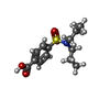 ChemComp-RTO: |
-Macromolecule #3: CHLORIDE ION
| Macromolecule | Name: CHLORIDE ION / type: ligand / ID: 3 / Number of copies: 1 / Formula: CL |
|---|---|
| Molecular weight | Theoretical: 35.453 Da |
-Experimental details
-Structure determination
| Method | cryo EM |
|---|---|
 Processing Processing | single particle reconstruction |
| Aggregation state | particle |
-Sample preparation
| Concentration | 15 mg/mL | |||||||||
|---|---|---|---|---|---|---|---|---|---|---|
| Buffer | pH: 7.5 Component:
Details: 20 mM HEPES, 200 mM NaCl, 0.0015% LMNG, 0.00015% CHS | |||||||||
| Grid | Model: Quantifoil R1.2/1.3 / Material: GOLD / Mesh: 300 / Support film - Material: CARBON / Support film - topology: HOLEY / Pretreatment - Type: GLOW DISCHARGE / Pretreatment - Time: 30 sec. / Pretreatment - Pressure: 0.025 kPa | |||||||||
| Vitrification | Cryogen name: ETHANE / Chamber humidity: 100 % / Chamber temperature: 283 K / Instrument: FEI VITROBOT MARK IV | |||||||||
| Details | this sample is a complex formed by composition of human OAT1, Fab targetting C-terminus of HsOAT1, and the inhibitor probenecid. |
- Electron microscopy
| Microscope | TFS KRIOS |
|---|---|
| Image recording | Film or detector model: FEI FALCON IV (4k x 4k) / Digitization - Dimensions - Width: 4096 pixel / Digitization - Dimensions - Height: 4096 pixel / Number grids imaged: 2 / Number real images: 22975 / Average electron dose: 52.0 e/Å2 |
| Electron beam | Acceleration voltage: 300 kV / Electron source:  FIELD EMISSION GUN FIELD EMISSION GUN |
| Electron optics | Illumination mode: OTHER / Imaging mode: BRIGHT FIELD / Cs: 2.7 mm / Nominal defocus max: 2.0 µm / Nominal defocus min: 1.0 µm / Nominal magnification: 96000 |
| Sample stage | Specimen holder model: FEI TITAN KRIOS AUTOGRID HOLDER / Cooling holder cryogen: NITROGEN |
| Experimental equipment |  Model: Titan Krios / Image courtesy: FEI Company |


